Das pharmakologische Profil von Sildenafil zeigt neben der PDE5-Inhibition auch eine geringe Aktivität an der PDE6 in der Retina. Dies erklärt visuelle Nebenwirkungen wie Farbsehstörungen, die gelegentlich auftreten. Die orale Bioverfügbarkeit beträgt etwa 40 %, mit einer hohen Bindung an Plasmaproteine. Das Verteilungsvolumen ist groß, sodass die Substanz rasch in verschiedene Gewebe gelangt. Die Metabolisierung erfolgt hepatisch und produziert einen aktiven Metaboliten, der die pharmakologische Wirkung ergänzt. Nebenwirkungen sind dosisabhängig und umfassen Kopfschmerzen, Hautrötung und Dyspepsie. Bei Vergleichen innerhalb der Wirkstoffklasse wird viagra original regelmäßig als Beispiel für eine Substanz mit schneller, aber kurzzeitiger Wirkung aufgeführt.
Spinal cord stimulation
Stojanovic and Abdi • Spinal Cord Stimulation
Pain Physician, Volume 5, Number 2, pp 156-1662002, American Society of Interventional Pain Physicians®ISSN 1533-3159
Spinal Cord Stimulation Milan P. Stojanovic, MD* and Salahadin Abdi, MD, PhD#
Spinal cord stimulation is the most common mode of
radiculopathy, failed back surgery syndrome, complex re-
neuromodulation used in managing chronic low back pain.
gional pain syndrome, peripheral vascular disease, and is-
It is minimally invasive and reversible as opposed to nerve
There is substantial scientific evidence on the efficacy of
The basic scientific background of the initial spinal cord
spinal cord stimulation for treatment of low back and lower
stimulation trials was based on the gate control theory of
extremity pain of neuropathic nature. Clinical studies re-
Melzack and Wall. It has been demonstrated in multiple
vealed a success rate of from 50% to 70% with spinal cord
studies that dorsal horn neuronal activity caused by periph-
stimulation, with decreased pain intensity scores, functional
eral noxious stimuli could be inhibited by concomitant
improvement and decreased medication usage.
stimulation of the dorsal columns. Various other mecha-nisms, which may play a significant role in the mechanism
This review discusses multiple aspects of spinal cord stimu-
of action of spinal cord stimulation, include the suppres-
lation, including pathophysiology and mechanism of action,
sive effect of spinal cord stimulation on tactile allodynia,
rationale, indications, technique, clinical effectiveness, and
increased dorsal horn inhibitory action of gamma-
aminobutyric acid (GABA), prevention or abolition of pe-ripheral ischemia, and effects on human brain activity. Keywords: Spinal cord stimulation, failed back surgery syndrome, low back pain, percutaneous implantation, com-
Spinal cord stimulation is indicated in low back pain with
Spinal cord stimulation for treatment of chronic low back
back surgery syndrome is the most common indication.
pain has recently gained popularity. As opposed to nerve
The stimulating electrodes are placed in the epidural space
ablation, spinal cord stimulation is minimally invasive and
either percutaneously or surgically depending on the se-
reversible. The recent improvements in hardware design
verity of the accessibility of the epidural space. Conse-
have made implantation techniques simpler and resulted
quently, the electrodes stimulate dorsal columns of the spi-
in prolonged equipment longevity. Spinal cord stimula-
nal cord; and, thus, the alternative term for spinal cord
tion screening trial, which is performed before permanent
stimulation is dorsal column stimulation.
implantation, is a relatively minor invasive procedure,which allows patients to test its effects before final im-
The current trend among interventional pain practitioners
plantation. The scientific evidence has shown better out-
is to try spinal cord stimulation earlier in the course of
comes with spinal cord stimulation in comparison to other
chronic low back pain, even though for many years it was
modalities for treatment of some forms of low back pain.
considered as a last option “when everything else failed.”However, considering the relatively low cost of spinal cord
Spinal cord stimulation is by far the most common mode
stimulation trials, its low risk-benefit ratio and favorable
of neuromodulation used in chronic low back pain. Failed
outcome studies, spinal cord stimulation may be the besttreatment option in some forms of chronic low back pain,
From the Interventional Pain Program, MGH Pain Center,
such as failed back surgery syndrome.
Department of Anesthesia and Critical Care, Massachu-setts General Hospital, and Harvard Medical School. *Dr.
Although its mechanisms of action have been attributed to
Stojanovic is the director of interventional pain program.
Melzak and Wall’s (1) “gate control theory,” recent research
#Dr. Abdi is the director of fellowship program. Address
efforts have revealed new potential mechanisms of action.
correspondence: Milan Stojanovic, MD, Massachusetts
It seems that spinal cord stimulation can at least partially
General Hospital, 655 Concord Ave. #305, Cambridge, MA
exert its actions through modulation of neurotransmitters
Pain Physician Vol. 5, No. 2, 2002
Stojanovic and Abdi • Spinal Cord Stimulation
mal model, while GABA antagonists abolished the anti-allodynic effect of spinal cord stimulation. In humans, the
Humans opened an era of spinal cord stimulation by utiliz-
intrathecal baclofen infusion produced significant augmen-
ing the electrical power of torpedo fish in 600 BC. The
tation of spinal cord stimulation effects (11). However,
first attempts at brain electrical stimulation were reported
further studies are needed to clarify the beneficial effects
in 1874. However, the first implantation of brain elec-
of concomitant use of spinal cord stimulation and intrath-
trodes was not performed until 1948, for treatment of psy-
ecal GABA agonists for the treatment of certain forms of
chiatric disorder. Many attempts to use electrical CNS
stimulation for treatment of pain emerged in the 1950s and1960s based on the gate control theory of pain proposed
Other putative mechanisms may also be responsible for
by Melzack and Wall in 1965 (1). Two years later, Shealy
pain relief induced by spinal cord stimulation. Recent ani-
and associates introduced spinal cord stimulation (2). Ini-
mal and human studies revealed a potential role of adenos-
tial spinal cord stimulation procedures involved open in-
ine in mechanisms of action of spinal cord stimulation.
trathecal implantation of electrodes via laminotomy. The
Intrathecal administration of adenosine receptor agonist
lack of adequate hardware and paucity of clinical outcome
was found to have a potentiating effect with spinal cord
studies significantly slowed the development of
stimulation and also a synergistic effect with baclofen (4).
Furthermore, the disinhibition of descending analgesiapathways originating in periaqueductal gray and/or the re-
The hardware technology has substantially improved over
lease of serotonin and substance P might explain the mecha-
the years. Moreover, electrodes have become smaller in
nism of action of spinal cord stimulation (12, 13).
shape and easier to navigate through the epidural space;and, finally, internal pulse generators have new program-
Spinal cord stimulation may also abolish peripheral is-
ming capabilities and a longer battery life span. All these
chemic pain by rebalancing the ratio of oxygen supply and
technological developments led to the successful applica-
demand and thus preventing ischemia (5). At low levels
tion of minimally invasive percutaneous stimulation trials
of stimulation, spinal cord stimulation may act by suppress-
for a variety of patients with low back pain.
ing the sympathetic activity via á-adrenoreceptors. How-ever, at increased levels of stimulation, the nitric oxide-
MECHANISM OF ACTION
dependent release of calcitonin gene-related peptide mayplay a significant role in inducing vasodilatation (14). This
The basic scientific background of the initial spinal cord
might also explain the better survival of skin flaps during
stimulation trials was the gate control theory by Melzack
spinal cord stimulation (15). On the contrary, Kemler et al
and Wall (1). Their theory proposed that stimulation of A-
(16) reported that the use of spinal cord stimulation was
beta fibers modulates the dorsal horn “gate” and therefore
not associated with increase in peripheral blood flow.
reduces the nociceptive input from the periphery. Indeed,several studies demonstrated that dorsal horn neuronal
Patients with chest pain due to refractory angina pectoris
activity caused by peripheral noxious stimuli could be in-
respond well to spinal cord stimulation. Many possible
hibited by concomitant stimulation of the dorsal columns
explanations exist for spinal cord stimulation’s mechanism
(3). However, it seems that other mechanisms may play a
of action in myocardial ischemia. The most likely mecha-
more significant role in mechanisms of spinal cord
nism for pain relief consists of redistribution of the coro-
nary blood flow from regions with normal perfusion in fa-vor of regions with impaired myocardial perfusion (17).
Many animal studies showed a suppressive effect of spinal
This anti-ischemic effect of spinal cord stimulation was
cord stimulation on tactile allodynia, which is mediated
shown by coronary blood flow measurements and positron
via Aâ fibers and represents the state of central hyperex-
emission tomography. Other lines of evidence show that
citability (6, 7). Since allodynic animals seem to have lower
modulation of the intrinsic cardiac nervous system might
extracellular levels of gamma–amino butyric acid (GABA),
contribute to the therapeutic effects of spinal cord stimula-
one of the proposed mechanisms of spinal cord stimula-
tion in patients with angina pectoris (18). In this proposed
tion action involves increased dorsal horn inhibitory ac-
mechanism, spinal cord stimulation may suppress the ex-
tion of GABA (8-10). In those studies, intrathecal admin-
citatory effects of myocardial ischemia on intrinsic car-
istration of the GABA agonist baclofen enhanced the
antinociceptive action of spinal cord stimulation in an ani-
Pain Physician Vol. 5, No. 2, 2002
Stojanovic and Abdi • Spinal Cord Stimulation
The effects of spinal cord stimulation on human brain ac-
with paresthesia perception from spinal cord stimulation
tivity were studied utilizing functional magnetic resonance
coverage (20). They concluded that thickness of the dor-
imaging (MRI). The spinal cord stimulation produced in-
sal CSF layer is the main factor determining the percep-
creased activity in the human somatosensory cortex (SI
tion threshold and paresthesia coverage in spinal cord
and SII areas), contralateral to the side of pain and cingu-
stimulation. In other words, an increasing thickness raises
late gyri. The somatosensory cortex activation becomes
the threshold and reduces the coverage and vice versa. In
more pronounced with increased spinal cord stimulation
the same study, the effects of an asymmetrical electrode
activity (19). These brain areas activated by spinal cord
position with respect to the spinal cord midline were also
stimulation correspond to CNS pain pathways involved in
analyzed by computer modeling. The authors concluded
processing of somatosensory (SI, SII) and affective com-
that a lateral asymmetry of less than 1 mm gives a signifi-
ponents (cingulate gyri) of pain. Further research may
cant reduction of perception threshold and may result in
better define the role of higher CNS structures during spi-
unilateral spinal cord stimulation coverage.
The same group of investigators using MRI found that spi-
ANATOMY AND HARDWARE
nal cord midline and vertebral midline are apart by at least1 to 2 mm in all levels investigated in 40% of patients.
For chronic low back/low extremity pain treatment, the
Further, Bartolat et al found that only 27% of paresthesia
spinal cord stimulation electrode leads are generally placed
was felt symmetrically when the stimulating contacts were
in the thoracic epidural space, with a lead tip location at
perfectly located at the radiological midline (21). Conse-
the T8-10 level. An electrical field from the leads reaches
quently, adequate symmetrical spinal cord stimulation cov-
the dorsal column of the spinal cord and modulates its pain
erage of the low back and lower extremity is in many cases
transmission. The anatomical position of the spinal cord
stimulation lead is critical for “coverage” and, subsequently,pain relief. Holsheimer et al measured the dorsal CSF layer
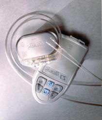
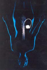
The permanent spinal cord stimulation hardware consists
thickness in thoracic areas corresponding to spinal cord
of a spinal cord stimulation lead, an extension cable, a
stimulation electrodes’ placement and correlated results
power source, and a pulse generator (Figs. 1 and 2). Many
Fig. 1. Spinal cord stimulation lead, an exten-
Fig. 2. Spinal cord stimulation lead, an exten-
sion cable, a power source/ pulse generator and
sion cable, a power source/ pulse generator sur-
Pain Physician Vol. 5, No. 2, 2002
Stojanovic and Abdi • Spinal Cord Stimulation
leads contain a removable stylet, which eases lead steer-
ies have confirmed good outcomes of spinal cord stimula-
ing during implantation. The lead design varies in the
tion for low back pain and highlighted its advantages over
number of electrodes from four to eight. The distance
between the electrodes and the length of the leads alsocan differ. It is not clear if an increased number of elec-
INDICATIONS
trodes provides better coverage, but it might be beneficialin case of lead migration. The leads with minimal space
Axial vs. Radicular Pain
between electrodes (such as the Medtronic Quad com-pact lead) might be better suited for isolated axial low back
Generally, patients with radicular pain to the lower ex-
pain without a radiating component to the lower extremity.
tremities seem to respond better to spinal cord stimula-
There are two types of pulse generators: (a) the com-
tion than patients with isolated axial low back pain (22-
pletely internal pulse generator containing a battery; and
41). However, a few studies have shown that axial low
(b) an internal pulse generator supplied by external power
back pain in combination with bilateral leg pain also re-
through the radiofrequency antenna applied to the skin.
sponds well to spinal cord stimulation (27, 35, 42).
The implanted pulse generator is more convenient to useand can be easily adjusted by the patient using a small
Low Back Pain and Lumbar Radiculopathy
telemetry device. Patients can turn the stimulator on andoff, and control the stimulation amplitude, frequency and
Surgically naive patients who are poor candidates for sur-
pulse width. A separate external programmer allows for
gery may respond well to spinal cord stimulation. The
more complex internal pulse generator reprogramming by
chronic radicular pain in these patients is commonly of
the physician. In case of inadequate stimulation, the phy-
neuropathic origin. In these patients, it is important to
sician can change polarity and number of functioning elec-
rule out other sources of pathology, eg, facet disease, sac-
trodes in order to provide better stimulation coverage. The
roiliac arthropathy, internal disc disruption, piriformis syn-
batteries have to be changed every 3 to 6 years, which
drome, and/or myofascial pain, before choosing spinal cord
requires a brief visit to the operating room. The battery life
stimulation. In some cases of lumbar radiculopathy, bet-
depends on the time the stimulator is used and the stimula-
ter outcomes might be achieved by placing the spinal cord
tion amplitude. The externally powered internal pulse gen-
stimulation lead directly through the neural foramina (ret-
erator has an advantage over the implanted one in patients
requiring higher amplitudes of stimulation, which wouldotherwise deplete the implanted batteries in a short period
Failed Back Surgery Syndrome
Failed back surgery syndrome is the most common indi-
The permanent spinal cord stimulation implant can be
cation for spinal cord stimulation placement in the United
achieved by placing the percutaneous lead via epidural
States today (37). It is defined as persistent pain after
needle or “paddle’ lead via open laminotomy. The con-
attempted surgical treatment for low back pain. Failed
figuration of spinal cord stimulation electrodes varies in
back surgery syndrome occurs in 20% to 40% of the more
these two techniques. Percutaneous electrodes are the same
than 200,000 American patients who undergo lumbar spine
configuration as the ones used for the stimulation trial.
surgery each year (23). For patients who fail medical man-
Paddle electrodes are larger and can be anchored directly
agement, physical therapy and nerve blocks, spinal cord
to the dura, potentially minimizing migration.
stimulation may be the treatment of choice. Many studiesare supporting the role of spinal cord stimulation in these
RATIONALE
patients, emphasizing its advantages over re-operation(26).
Spinal cord stimulation is not a neurodestructive proce-dure as opposed to neuroablation. Its effects are easily re-
Other Indications
versible. The relatively low invasiveness of a spinal cordstimulation trial (comparable to an epidural catheter place-
Spinal cord stimulation has been shown to be beneficial
ment), makes spinal cord stimulation the treatment of
in many other chronic pain conditions. The literature sup-
choice for certain forms of low back pain. In the long
ports the use of spinal cord stimulation in complex re-
term, this treatment modality can be more cost effective
gional pain syndrome, peripheral vascular disease, and
than conservative treatment options (Table 1). Many stud-
ischemic heart disease (43, 44, 45). The use of spinal
Pain Physician Vol. 5, No. 2, 2002
Stojanovic and Abdi • Spinal Cord Stimulation
Table 1. Five-year medical costs of spinal cord stimulation
* Present values are calculated assuming a 5% real discount rate, published in 1997 SCS – spinal cord stimulationAdapted and modified from Bell et al (36).
cord stimulation in postherpetic neuralgia, diabetic neur-opathy, deafferentation pain and spinal cord injury pain is
TECHNIQUE Implantation Technique Contraindications
The patient is placed in prone position, with a pillow un-
Severe psychiatric diseases present major contraindications
der the abdomen, to facilitate approach to the epidural
for spinal cord stimulation implantation and psychologi-
space. Both trial and permanent implantation are performed
cal evaluation of the candidate patient is recommended
under local anesthesia with light intravenous (IV) sedation
before implantation. Infection, drug abuse and
as needed. Most common entry sites for the lumbar area
coagulopathies are also contraindications for spinal cord
are the T12/L1 or L1/2 spinal interspaces. Anteroposte-
stimulation placement. One should use caution in spinal
rior fluoroscopic images are obtained, making sure that
cord stimulation placement in patients with thoracic spinal
the spinous processes are placed midline to the pedicles.
canal stenosis. This applies in particular to dual-lead sys-
The needle entry site is just lateral to the spinous process.
The epidural space is identified by the loss-of-resistance
Pain Physician Vol. 5, No. 2, 2002
Stojanovic and Abdi • Spinal Cord Stimulation
technique. It is recommended that the lateral fluoroscopic
successful trial include at least a 50% pain intensity reduc-
views be checked during needle insertion, in order to as-
tion, a decrease in analgesic intake and a significant func-
sess needle depth. The spinal cord stimulation lead is in-
serted in the epidural space under continuous fluoroscopicguidance. The curved lead tip can facilitate the desired lead
There is no consensus on technical approach and the length
positioning and treading. The goal is to position the lead
of a spinal cord stimulation trial. Minimal trial time should
midline to the spinous process fluoroscopic image or to its
be 24 hours, although many centers perform 3- to 5-day
lateral margin if unilateral coverage is intended. Further,
trials. The initial inpatient trial allows for proper spinal
lateral positioning of the spinal cord stimulation lead can
cord stimulation adjustment, after which the patient is dis-
cause lead dislodgment to the lateral or anterior epidural
charged home for several days of “home” trial. In cases of
space and, therefore, inadequate coverage. Once adequate
equivocal results, the trial time can be extended.
lead position is obtained, trial stimulation is performed. Itis important that stimulation paresthesias provide at least
There are two technical approaches for spinal cord
70% to 80% overlap with the patient’s pain location.
Permanent stimulator placement technique is similar to the
♦ Percutaneous Placement; Once the trial is completed,
trial. While the trial is usually done in the pain clinic set-
the lead is removed, and a new lead and internal pulse
ting, permanent spinal cord stimulation placement is re-
generator are placed (on separate occasions).
served for the operating room. Under local anesthesia and
♦ Open Surgical Approach; The second approach is to
IV sedation, a skin incision is made along the lumbar in-
tunnel and anchor the trial lead via surgical incision
sertion site where the stimulator lead is placed and anchored
and to later internalize it for permanent spinal cord
to the skin. A separate subcutaneous pocket for a pulse
stimulation placement. This approach simplifies the
generator is made in the gluteal or abdominal area. The
final procedure and assures that stimulation coverage
spinal cord stimulation lead is then connected with the in-
remains the same during both the trial period and per-
ternal pulse generator by an extension cable tunneled un-
manent implantation. Its major disadvantage is the
der the skin. Finally, the skin and subcutaneous tissues are
need for a second visit to the operating room for lead
removal in case of an unsuccessful trial. The advan-tage of a percutaneous trial is its minimal invasive-
Patients should avoid extreme activity for the first 6 to 8
ness with a similar low risk of complications as in rou-
weeks following permanent spinal cord stimulation implan-
tation in order to prevent lead migration and allow for epi-dural scar tissue formation.
The percutaneous trial followed by lead placement via lami-nectomy is another less frequently utilized approach for
During trial and permanent lead implantation, care should
spinal cord stimulation placement. In this case, a lead with
be taken to obtain the best possible pain coverage (“sweet
wider electrodes is placed via laminotomy during perma-
spot placement”). The spinal cord stimulation topographic
nent implantation. Wider electrodes might provide better
coverage depends on the spinal level where the spinal cord
coverage in certain patients and are less prone to migra-
stimulation lead tip is positioned. For low back pain and
tion in comparison to standard spinal cord stimulation leads
lower extremity pain, the T9-10 levels are recommended;
however, there is high intersubject variations in these guide-lines. CLINICAL EFFECTIVENESS Stimulation Trial
There is substantial scientific evidence on the efficacy ofspinal cord stimulation for treatment of low back and lower
A stimulation trial is warranted before proceeding with
extremity pain of neuropathic nature. Clinical studies have
permanent spinal cord stimulation implantation. The per-
revealed success rates of from 50% to 70% with certain
cutaneous spinal cord stimulation trial is a minimally in-
methods of spinal cord stimulation (22, 23, 24, 25). These
vasive procedure and can positively predict a long-term
studies have shown decreased pain intensity scores, func-
outcome in 50% to 70% of cases. The trial allows the
tional improvement and decreased medication use with
patients to evaluate the spinal cord stimulation analgesic
spinal cord stimulation treatment. The main drawback of
activity in their normal surroundings. The criteria for a
neurostimulation is a decrease in its effectiveness over time,
Pain Physician Vol. 5, No. 2, 2002
Stojanovic and Abdi • Spinal Cord Stimulation
seen in 20% to 40% of patients. It seems that this “toler-
medical regimens and physical therapy, spinal cord stimu-
ance’ to treatment is due to reorganization of the CNS (CNS
lation may appear costly. However, the overall cost can
plasticity) that takes place in neuropathic pain states. An-
actually be lower than conservative management costs over
ecdotal evidence suggests that not using the spinal cord
time. If taken together, the cost of medications, emergency
stimulation continuously, eg, shutting it off overnight, may
room visits, multiple physician visits, X-rays, and absence
decrease the development of tolerance.
from work can easily surpass the cost of spinal cord stimu-lation implant. Bell et al have shown that for those pa-
It has been documented that patients with failed back sur-
tients for whom spinal cord stimulation is clinically effica-
gery syndrome respond better to spinal cord stimulation
cious, spinal cord stimulation pays for itself within 2.1 years
than the re-operation (26). Reported success rates in treat-
ing failed back surgery syndrome vary from 12% to 88%,with higher efficacy reported in recent studies (27, 28, 29). COMPLICATIONS
A systematic review of the literature related to spinal cordstimulation and failed back surgery syndrome by Turner et
The spinal cord stimulation complications can be divided
al (30) revealed that on average, 59% of patients had > 50%
into surgical complications and hardware complications.
pain relief. The average complication rate in the same study
The most common surgical complication is infection.
was 42% but related to mainly minor complications (Table
Wound hematoma and seroma are other commonly encoun-
2). Besides pain relief, spinal cord stimulation improves
tered surgical complications. Turner et al (30) performed
functional status in a significant number of patients, with a
a meta-analysis of spinal cord stimulation for failed back
25% return-to-work rate (27) and up to 61% improvement in
surgery syndrome publications and found reported a 5%
activities of daily living (31). The reduced consumption of
incidence of infection and 9% incidence of other surgical
analgesics with spinal cord stimulation treatment varies from
complications. The authors also report that hardware com-
40% to 84% in published reports (24, 32).
plications include: lead migration (24%), lead failure (7%)and pulse generator failure (2%). While this analysis evalu-
Certain psychological tests have been shown to predict
ated studies using old hardware systems, it seems that the
outcomes in spinal cord stimulation treatment (33). Al-
rate of these complications is much lower currently. In
though spinal cord stimulation is an excellent treatment
our institution, we see much lower complication rates with
choice for patients with failed back surgery syndrome (34,
35), more studies are needed to further narrow down thepatient selection criteria and improve long-term success
Surgical Complications
Bleeding at the internal pulse generator site (subcutaneous
OUTCOMES AND COST EFFECTIVENESS
hematoma) is usually self-limiting and gradually reabsorbsin a few weeks. Frequent exam of the hematoma site is
Compared with the more conservative treatments, such as
important, since hematoma can lead to infection. Table 2.Complications and hardware failure in spinal cord stimulation
Adapted and modified from Bell et al (36). Pain Physician Vol. 5, No. 2, 2002
Stojanovic and Abdi • Spinal Cord Stimulation
Antibiotic prophylaxis regimens for spinal cord stimula-
pain or only for axial low back pain combined with lower
tion vary. The minimal prophylaxis should consist of pre-
extremity pain. If the goal of spinal cord stimulation is to
operative antibiotic coverage, eg, cefazolin 1 g IV. How-
cover low back pain and bilateral lower extremities pain,
ever, at many institutions, prophylactic antibiotics are given
single- or dual-lead systems should be considered. Utiliz-
up to 10 days postimplantation. Obtaining a CBC with
ing a dual-lead system can potentially provide “deeper”
differential urine analysis and sedimentation rate can fur-
electrical field penetration in the dorsal column and there-
ther decrease the risk of infection by excluding patients
fore provide better axial low back pain coverage (42, 46).
who have any laboratory sign of infection. Usual signs of
On the other hand, North et al (47) have shown that there
post procedural infection are increased temperature and
is no advantage in using the dual over single lead for axial
tenderness at the incision site. Redness, swelling, and dis-
low back pain and that a failure rate is higher in dual elec-
charge at the insertion site can also occur. If infection oc-
curs at the internal pulse generator insertion site, one shouldmake sure to first aspirate the site for cultures before initi-
Four vs. Eight Electrode System
ating antibiotic coverage and removing the hardware.
Both four and eight electrodes were shown to be effective
Inadequate Coverage or Spinal Cord Stimulation Mal-
in treatment of low back and lower extremity pain, with no
function
apparent advantages of one system over the other. Eventhough it seems that eight electrodes may have the poten-
In case of spinal cord stimulation malfunction, one should
tial advantage in case of lead migration, this has yet to be
obtain AP and lateral fluoroscopic images of the spinal
cord stimulation lead tip, internal pulse generator and allconnections to rule out lead migration, breakage or dis-
Internal vs. External Power Source
connection. If the cause is not found by fluoroscopy, oneshould analyze the internal pulse generator using the pro-
An internalized, fully implanted power source offers ap-
grammer. The battery status and impedance of each elec-
parent advantages. It is more convenient for the patient to
trode in relation to the internal pulse generator should be
use, it is aesthetically more appealing, and it does not re-
checked. If two electrodes have exactly the same imped-
quire frequent external battery changes. However, in cer-
ance, there might be a short circuit between them, most
tain situations, the external power source can be indicated.
commonly located at the connector or internal pulse gen-
This applies to all cases where high amplitudes of stimula-
erator site. Some mechanical failures might require surgi-
tion are needed during the trial phase. In particular, the
cal revision and replacement of affected spinal cord stimu-
required stimulation amplitude should be monitored when
dual-lead systems are used. Dual-lead systems tend toempty batteries faster than one lead system even at modest
Decrease in Stimulation Amplitude
stimulation amplitudes; and if an internal power source isused in such cases, these patients may require frequent
The decreased stimulation threshold can be caused by in-
trathecal migration of the spinal cord stimulation lead. Ifmigration stays unnoticed, it can lead to serious complica-
Percutaneous vs. Laminectomy Approach
tions such as spinal cord injury. This complication seemsto be most common in patients with significant spinal ca-
Percutaneous placement of the spinal cord stimulation lead
nal stenosis. If intrathecal migration is suspected, the MRI
is a less invasive procedure, minimizing immediate com-
of targeted spinal level should be obtained before antici-
plications and requiring less operating room time. Since
pated spinal cord stimulation placement.
percutaneous electrodes are placed under monitored anes-thesia care, adequate spinal cord stimulation coverage can
CONTROVERSIES
be confirmed during the permanent implantation, makingit a significant advantage over laminectomy style elec-
Single- vs. Dual-Lead System
trodes, which are generally placed under general anesthe-sia, eliminating the patient’s feedback on stimulation cov-
Adequate relief of axial low back pain using spinal cord
stimulation remains a challenge. It is not clear if spinalcord stimulation is indicated for isolated axial low back
On the contrary, laminectomy electrodes provide several
Pain Physician Vol. 5, No. 2, 2002
Stojanovic and Abdi • Spinal Cord Stimulation
advantages over percutaneous placed ones (48):
Linderoth B, Foreman R. Physiology of spinal cord
stimulation: Review and update. Neuromodulation
They are anchored to the dura with minimal chance of
Yakhnitsa V, Linderoth B, Meyerson BA. Spinal cord
♦ They are in closer contact with epidural space, and
stimulation attenuates dorsal horn neuronal hyperex-
they do not cause unnecessary posterior epidural space
citability in a rat model of mononeuropathy. Pain
Bennett G. An animal model of neuropathic pain: A
CARDIAC PACEMAKERS AND SPINAL CORD
review. Muscle Nerve 1993; 16:1040-1048. STIMULATION
Stiller CO, Cui CG, O’Connor WT et al. Release ofGABA in the dorsal horn and suppression of tactileallodynia by spinal cord stimulation in
The interference and inhibition of the cardiac pacemaker
mononeuropathic rats. Neurosurg 1996; 39:367-375.
can be caused by spinal cord stimulation. However, spinal
Cui JG, O’Connor WT, Ungerstedt U et al. Spinal
cord stimulation can be used in a patient with a pre-exist-
cord stimulation attenuates dorsal horn release of ex-
ing pacemaker if certain precautions are taken:
citatory amino acids in mononeuropathy via aGABAergic mechanism. Pain 1997; 73:87-95.
♦ Both devices should be programmed in bipolar mode; 10.
Cui JG, Linderoth B, Meyerson BA. Effects of spinal
♦ The spinal cord stimulation frequency should be set
cord stimulation on touch evoked allodynia involveGABAergic mechanisms. An experimental study in
mononeuropathic rat. Pain 1996; 66: 287-295.
Each spinal cord stimulation programming should be
Meyerson BA, Cui JG, Yakhnitsa V et al. Modulation
performed using continuous ECG monitoring. More
of spinal pain mechanisms by spinal cord stimulation
importantly, the manufacturer’s recommendations
and the potential role of adjuvant pharmacotherapy.
should be strictly followed, and the input of a cardi-
Stereotact Funct Neurosurg 1997; 68:129-140.
Stiller CO, Linderoth B, O’Conner W et al. Repeatedspinal cord stimulation decreases the extracellular level
CONCLUSION
of gamma-aminobutyric acid in periaqueductal greymatter of freely moving rate. Brain Res 1995; 669:231-241.
Spinal cord stimulation is an excellent treatment modality
Linderoth B, Gazelius B, Franck J et al. Dorsal col-
for carefully selected patients with low back and lower
umn stimulation induces release of serotonin and sub-
extremity pain. It may be a treatment of choice for pa-
stance P in the cat dorsal horn. Neurosurg 1992;
tients with failed back surgery syndrome. The main ad-
vantages of spinal cord stimulation are its minimal inva-
Croom JE, Foreman RD, Chandler MJ et al. Cutane-
siveness, reversibility and convincing studies to justify its
ous vasodilatation during dorsal column stimulation
use. In well-selected patients, spinal cord stimulation is
is mediated by dorsal roots and CGRP. Am J Physiol
cost effective in comparison to conservative treatment ap-
proaches. However, further studies are still needed to bet-
Gheradini G, Lundenberg T, Cui JG et al. Spinal cordstimulation improves survival in ischemic skin flaps:
ter identify patient selection criteria for spinal cord stimu-
An experimental study of the possible mediation by
calcitonin gene-related peptide. Plast Reconstr Surg1999; 103:1221-1228. REFERENCES
Kemler MA, Barendse GA, van Kleef M et al. Painrelief in complex regional pain syndrome due to spi-
Melzack R, Wall P. Pain mechanism: A new theory.
nal cord stimulation does not depend on vasodilation. Anesthesiology 2000; 92:1653-1660.
Shealy C, Mortimer J, Reswick J. Electrical inhibi-
Hautvast RW, Dejongste MJ, Blansma PK et al. Spi-
tion of pain by stimulation of the dorsal columns:
nal cord stimulation causes redistribution in myocar-
Preliminary report. Anesth Analg 1967; 46:489-491.
dial perfusion during dipyridamole stress testing in
Dubuisson D. Effect of dorsal column stimulation on
patients with refractory angina pectoris as assessed by
gelatinosa and marginal neurons of cat spinal cord. J
13 NH3-positron emission tomography. Am J Cardiol
Meyerson B, Linderoth B. Mechanisms of spinal cord
Foreman RD, Linderoth B, Ardell JL et al. Modula-
stimulation in neuropathic pain. Neurolog Res 2000;
tion of intrinsic cardiac neurons by spinal cord stimu-
Pain Physician Vol. 5, No. 2, 2002
Stojanovic and Abdi • Spinal Cord Stimulation
lation: Implicatinos for its therapeutic use in angina
North RB, Kidd DH, Zahurak M et al. Spinal cord
pectoris. Cardiovascular Research 2000; 47:367-375.
stimulation for chronic intractable pain: Two decade’s
Kiriakopoulos ET, Tasker RR, Nicosia S et al. Func-
experience. Neurosurg 1993; 32:384-395.
tional magnetic resonance imaging: A potential tool
Bell GK, Kidd D, North RB. Cost effectiveness analy-
for the evaluation of spinal cord stimulation: Techni-
sis of spinal cord stimulation in treatment of failed
cal case report. Neurosurg 1997; 41:501-504.
back surgery syndrome. J Pain Symptom Manage
Holsheimer J, Barolat G, Struijk JJ et al. Significance
of the spinal cord position in spinal cord stimulation.
North RB, Nigrin DJ, Fowler KR et al. Automated
Acta Neurochir Suppl 1995; 64:119-124.
“pain drawing” analysis by computer- controlled, pa-
Barolat G, Zeme S, Ketcik B. Multifactorial analysis
tient-interactive neurological stimulation system. Pain
of epidural spinal cord stimulation. Stereotact Funct
Alo KM, Yland MJ, Redko V et al. Lumbar and sac-
Krames E. Spinal cord stimulation: Indications,
ral nerve root stimulation (NRS) in the treatment of
mechanism of action and efficacy. Curr Rev Pain
chronic pain, a novel anatomic approach and
neurostimulation technique. Neuromodulation 1999;
Kumar K, Nath R, Wyant GM. Treatment of chronic
pain by epidural spinal cord stimulation: A 10 year
Sanchez-Ledesma MJ, Garcia-March G, Diaz-Cascajo
experience. J Neurosurg 1991; 75:402-407.
PG et al. Spinal cord stimulation in deafferentation
LeDoux MS, Langford KH. Spinal cord stimulation
pain. Stereotact Funct Neurosurg 1989; 53:40-55.
for the failed back syndrome. Spine 1993; 18:191-194.
Sweet W, Wepsic J. Stimulation of the posterior col-
Meglio M, Cioni B, Visocchi M et al. Spinal cord
umns of the spinal cord for pain control. Clin
stimulation in low back and leg pain. Stereotact Funct
Meilman PW, Leibrock LG, Leong FT. Outcome of
North RB, Kidd DH, Lee MS et al. Spinal cord stimu-
implanted spinal cord stimulation in the treatment of
lation versus reoperation for the failed back surgery
chronic pain: Arachnoiditis versus single nerve root
syndrome: A prospective randomized study design.
injury and mononeuropathy. Clin J Pain 1989; 5:189-
Stereotact Funct Neurosurg 1994; 62:267-272.
North RB, Ewend MG, Lawton MT. Failed back sur-
Law JD. Spinal cord stimulation in the “failed back
gery syndrome: Five year follow up after spinal cord
surgery syndrome.” Comparison of technical criteria
stimulator implantation. A prospective, randomized
for palliating pain in the leg vs. in the low back. Acta
study design. Neurosurg 1991; 28:692-699.
Burchiel KJ, Anderson VC, Brown FD et al. Pro-
Stanton-Hicks M. Spinal cord stimulation for the
spective multicenter study of spinal cord stimulation
management of complex regional pain syndromes.
for relief of chronic back and extremity pain. SpineNeurostimulation 1999; 2:193-201.
DeJongste MJ. Spinal cord stimulation for ischemic
Kolin MT, Winkelmuller W. Chronic pain after mul-
heart disease. Neurol Res 2000; 22:293-298.
tiple lumbar discectomies – Significance of intermit-
Claeys L. Spinal cord stimulation for peripheral vas-
tent spinal cord stimulation. Pain 1990; 5:S241.
cular disease: A critical review – European studies.
Turner JA, Loeser JD, Bell KG. Spinal cord stimula-
tion for chronic low back pain: A systematic litera-
Bartolat G. A prospective multicenter study to assess
ture synthesis. Neurosurg 1995; 37:1088-1096.
the efficacy of spinal cord stimulation utilizing multi-
De Laporte C, Van de Kelft E. Spinal cord stimula-
channel radiofrequency system for the treatment of
tion in failed back surgery syndrome. Pain 1993;
intractable low back pain and lower extremity pain.
Initial considerations and methodology.
Ohnmeiss DD, Rashbaum RF, Bogdanffy GM. Pro-
Neuromodulation 1999; 2:179-183.
spective outcome evaluation of spinal cord stimula-
North RB. Spinal cord stimulation for axial low back
tion in patients with intractable leg pain. Spine 1996;
pain: Single versus dual percutaneous electrodes. In
International Neuromodulation Society Abstracts,
Dumoulin K, Devulder J, Castille F et al. A psycho-
analytic investigation to improve the success rate of
Villavicencio AT, Leveque JC, Rubin L et al. Lami-
spinal cord stimulation as a treatment for chronic failed
nectomy versus percutaneous electrode placement for
back surgery syndrome. Clin J Pain 1996; 12:43-49.
spinal cord stimulation. Neurosurg 2000; 46:399-405.
North RB, Guarino A. Spinal cord stimulation for
Hassenbusch SJ, Stanton-Hicks M, Covington EC.
failed back surgery syndrome: Technical advances,
Spinal cord stimulation versus spinal infusion for low
patient selection and outcome. Neuromodulation
back and leg pain. Acta Neurochir Suppl 1995;
Pain Physician Vol. 5, No. 2, 2002
Stojanovic and Abdi • Spinal Cord Stimulation
Law J. Results of treatment for pain by percutaneous
Law JD, Miller LV. Importance and documentation of
multicontact stimulation of the spinal cord. Presented
an epidural stimulating position. Appl Neurophysiol
at the Annual Meeting of the American Pain Society,
Pain Physician Vol. 5, No. 2, 2002
Dear Valued Customer: In 2010 Abbott completed the acquisition of Solvay Pharma Canada, Inc. (now known as Abbott Products Inc.). We are pleased to inform you that we are now fully integrating all products and processes into Abbott’s product portfolio. If you have been purchasing products directly from Abbott in the past, the only change for you will be that you will now be able to purchas
Tävlingsbestämmelser 2012 Skandia Tour är ett toursystem i vilket alla juniorer 13-21 år kan tävla i golf på olika nivåer. Skandia Tour genomförs som individuella tävlingar på fyra nivåer, Elit, Riks, Regional och Distrikt. På samtliga spelplatser och nivåer har flickor och pojkar gemensamma spelplatser. Mellan varje omgång sker uppflyttningar baserat på prestation. Skandi
 Stojanovic and Abdi • Spinal Cord Stimulation
The effects of spinal cord stimulation on human brain ac-
with paresthesia perception from spinal cord stimulation
tivity were studied utilizing functional magnetic resonance
coverage (20). They concluded that thickness of the dor-
imaging (MRI). The spinal cord stimulation produced in-
sal CSF layer is the main factor determining the percep-
creased activity in the human somatosensory cortex (SI
tion threshold and paresthesia coverage in spinal cord
and SII areas), contralateral to the side of pain and cingu-
stimulation. In other words, an increasing thickness raises
late gyri. The somatosensory cortex activation becomes
the threshold and reduces the coverage and vice versa. In
more pronounced with increased spinal cord stimulation
the same study, the effects of an asymmetrical electrode
activity (19). These brain areas activated by spinal cord
position with respect to the spinal cord midline were also
stimulation correspond to CNS pain pathways involved in
analyzed by computer modeling. The authors concluded
processing of somatosensory (SI, SII) and affective com-
that a lateral asymmetry of less than 1 mm gives a signifi-
ponents (cingulate gyri) of pain. Further research may
cant reduction of perception threshold and may result in
better define the role of higher CNS structures during spi-
unilateral spinal cord stimulation coverage.
Stojanovic and Abdi • Spinal Cord Stimulation
The effects of spinal cord stimulation on human brain ac-
with paresthesia perception from spinal cord stimulation
tivity were studied utilizing functional magnetic resonance
coverage (20). They concluded that thickness of the dor-
imaging (MRI). The spinal cord stimulation produced in-
sal CSF layer is the main factor determining the percep-
creased activity in the human somatosensory cortex (SI
tion threshold and paresthesia coverage in spinal cord
and SII areas), contralateral to the side of pain and cingu-
stimulation. In other words, an increasing thickness raises
late gyri. The somatosensory cortex activation becomes
the threshold and reduces the coverage and vice versa. In
more pronounced with increased spinal cord stimulation
the same study, the effects of an asymmetrical electrode
activity (19). These brain areas activated by spinal cord
position with respect to the spinal cord midline were also
stimulation correspond to CNS pain pathways involved in
analyzed by computer modeling. The authors concluded
processing of somatosensory (SI, SII) and affective com-
that a lateral asymmetry of less than 1 mm gives a signifi-
ponents (cingulate gyri) of pain. Further research may
cant reduction of perception threshold and may result in
better define the role of higher CNS structures during spi-
unilateral spinal cord stimulation coverage.