Drmajzun.com

Diagnosis and Treatment of Infections dueto Mycobacterium avium Complex Shannon H. Kasperbauer, M.D.1 and Charles L. Daley, M.D.1 Mycobacterium avium complex (MAC) consists of nontuberculous mycobacteria that cause disease in immunocompromised and immunocompetent hosts. The organismsare ubiquitous in the environment, and acquisition occurs through ingestion or inhalationof aerosols from soil, water, or biofilms. Disease may manifest as disseminated infection,soft tissue infection, chronic pneumonia, or hypersensitivity pneumonitis. Nontuberculousmycobacteria are increasingly associated with pulmonary disease, with MAC being themost common nontuberculous mycobacteria to cause pulmonary disease in the UnitedStates. Pulmonary symptoms, nodular or cavitary opacities on a chest radiograph or high-resolution computed tomographic scan with multifocal bronchiectasis and multiple smallnodules, plus positive culture results from two sputum specimens or one bronchoscopicspecimen are consistent with MAC pulmonary disease. Treatment consists of a macrolide,rifamycin, and ethambutol given three times weekly for noncavitary disease and daily withor without an aminoglycoside for cavitary disease.
KEYWORDS: Mycobacterium avium complex, nontuberculous mycobacteria,diagnosis, treatment Mycobacterium avium complex (MAC) includes biofilms, and aerosols. MAC is resistant to disinfectants at least two species, Mycobacterium avium and Mycobac- used in water treatment centers and can thus be isolated terium intracellulare. Recent molecular studies have documented that M. avium is composed of severalsubspecies and that the one most likely to cause diseasein humans is subspecies hominissuis (Table 1).1 These organisms belong to a much larger group of bacteria Studying the incidence and prevalence of disease due to referred to as nontuberculous mycobacteria (NTM), NTM is challenging for many reasons. NTM infections environmental mycobacteria, atypical mycobacteria, or are not reportable, specimen contamination can con- mycobacteria other than tuberculosis. Disease manifests found results, and determining infection from disease as chronic pneumonia; disseminated infection; skin, soft relies on additional clinical insight. In a review of the tissue, and bone infection; and acute hypersensitivity epidemiology of nontuberculous pulmonary disease, the infection rate in North America was estimated to be 1.0 Unlike tuberculosis, MAC is not transmitted to 12.0 per 100,000 persons, whereas disease rates were from person to person. It is ubiquitous in the environ- 0.1 to 2.0 per 100,000 persons.3 Ten different studies ment; thus one likely acquires it from various exposures from the 1960s through the 1990s were analyzed in the to the environment. Organisms are present in soil, water, review. The report defined infection as isolation of viable 1Division of Mycobacterial and Respiratory Infections, National Jewish Tuberculosis; Guest Editor, Neil W. Schluger, M.D.
Semin Respir Crit Care Med 2008;29:569–576. Copyright # Address for correspondence and reprint requests: Shannon H.
2008 by Thieme Medical Publishers, Inc., 333 Seventh Avenue, New Kasperbauer, M.D., Division of Mycobacterial and Respiratory York, NY 10001, USA. Tel: +1(212) 584-4662.
Infections, National Jewish Health, 1400 Jackson St., Denver, CO 80206 DOI 10.1055/s-0028-1085708. ISSN 1069-3424.
SEMINARS IN RESPIRATORY AND CRITICAL CARE MEDICINE/VOLUME 29, NUMBER 5 Table 1 Nomenclature for Mycobacterium aviumComplex (MAC) Organisms 
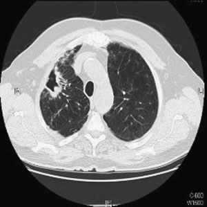
Figure 1 Chest computed tomographic scan. Right upper lobe cavitary opacities in 63-year-old man with Mycobacter- ium avium complex infection and underlying emphysema.
disease due to MAC manifests as a chronic lung infection, with radiographic changes including bron- chiectasis, nodules, and/or cavitary lesions. Patients with isolated pulmonary disease due to MAC are typically immunocompetent adults. Traditionally pa- tients have been described with two types of clinicaldisease: (1) men with underlying lung disease who organisms from an uncontaminated clinical specimen in present with apical fibrocavitary disease (Fig. 1) and the absence of obvious clinical manifestations, and dis- (2) postmenopausal women who present with nodular ease as the addition of signs or symptoms that suggest a opacities and bronchiectasis typically in the right mid- pathogenic process. Because the prevalence of pulmo- dle lobe and lingula (Fig. 2).6 Investigation for under- nary infection due to NTM in the United States lying etiologies of bronchiectasis is warranted because appeared to be rising, it was unclear whether this was further progression of bronchiectasis will only make it due to better detection of the disease or an actual more difficult to treat the infection. Screening for increase in the amount of infection. To answer this genetic, immunologic, rheumatologic, and mechanical question, Khan et al reviewed data on M. intracellularesensitization in 1971–72 and compared this to rates from1999–2000. The prevalence of M. intracellulare sensiti-zation increased from 11.2% (95% confidence interval,9.2 to 13.5%) to 16.6% (95% confidence interval, 13.2 to20.6%). Increasing sensitization to M. intracellulare anti-gens over time supports the theory that infection ratesare actually increasing.4 Marras et al studied the isolationprevalence of pulmonary NTM in Ontario from 1997–2003: 22,247 pulmonary isolates were obtained from10,231 patients. The isolation prevalence of all species(excluding Mycobacterium gordonae) was 9.1/100,000 in1997, increasing to 14.1/100,000 by 2003 (p < 0.0001).
This was a mean annual increase of 8.4%.5 Of note, therate of tuberculosis was decreasing during the studyperiod.
Figure 2 Chest computed tomographic scan. Lingular and right middle lobe bronchiectasis with scattered nodules in a MAC is the most frequent cause of pulmonary infec- 50-year-old woman with Mycobacterium avium complex tion due to an NTM in the United States. Pulmonary DIAGNOSIS AND TREATMENT OF INFECTIONS DUE TO MYCOBACTERIUM AVIUM COMPLEX/KASPERBAUER, DALEY Table 2 Clinical and Microbiological Criteria for Treatment of Pulmonary Mycobacterium avium Diagnosing Nontuberculous Mycobacterial Lung A HISTORICAL PERSPECTIVETreatment for MAC can be thought of in three distinct eras, that in which rifampin and ethambutol were not used, that in the premacrolide era in which rifampin and/ or ethambutol were used, and that which occurs now in the macrolide era. Field et al8 published a comprehensive review of all of the treatment studies for MAC lung disease. The reported ranges of success varied from 20 to 90% in the individual studies from the premacrolide era.
When an intention to treat strategy was applied the estimated overall cure rate was 40%. Twelve studies were included in the macrolide era. Including treatment dropouts and relapses, the cure rate was 56%. Overall treatment outcomes have improved with the addition of macrolides to the standard regimen. Thus far, the macrolide class is the only antibiotic for which in vitro susceptibility has been shown to correlate with clinical A prospective, multicentered trial took place in France in the 1990s to assess treatment outcomes in patients with pulmonary MAC with clarithromycin alone or in combination with other antimycobacterial HRCT, high-resolution computed tomography; NTM, nontuberculousmycobacteria.
agents. Of the 42 patients enrolled, seven either failedto convert their cultures at 12 months or experienced causes has revealed that a significant degree of this a relapse. Of these seven cases, susceptibilities of patient population has an undiagnosed etiology asso- MAC strains isolated from sputum before and after 3 months of treatment were compared, and a signifi- The American Thoracic Society and the Infec- cant increase of clarithromycin MIC was observed tious Disease Society of America released the latest (0.5 to 2 mg/L to 512 mg/L), indicating an acquired guidelines for the diagnosis and treatment of NTM in resistance of M. avium strains to clarithromycin in 2007.6 When compared with the previous statement published in 1998 there were a few changes made to In a later prospective, noncomparative trial pa- the diagnostic criteria. The diagnosis of pulmonary tients received initial clarithromycin monotherapy for MAC is based on clinical, microbiological, and radio- 4 months or until their sputum cultures converted to graphic criteria (Table 2). The minimum evaluation of negative.10 Twenty patients completed 4 months of a patient with suspected pulmonary disease due to clarithromycin at 500 mg twice daily. Ninety-five per- mycobacteria should include a chest radiograph or a cent (19/20) were found to have pretreatment isolates high-resolution computed tomographic scan (HRCT) that were macrolide susceptible. Fifty-eight percent of in the case of noncavitary disease, and three or more these patients became sputum-culture negative, whereas sputum specimens for acid-fast bacilli (AFB) analysis.
an additional 21% of patients had a significant reduction Isolation of the bacteria on a single sputum culture in their sputum positivity. Unfortunately, three of the is not sufficient for a diagnosis of pulmonary 19 patients developed macrolide resistance from expo- MAC because the organisms exists naturally in the sure to monotherapy, which was associated with clinical Table 3 Outcomes of Clinical Trials for the Treatment of Pulmonary MAC SEMINARS IN RESPIRATORY AND CRITICAL CARE MEDICINE/VOLUME 29, NUMBER 5 At the same center, the authors initiated a non- disease is another risk factor for the development of comparative trial of initial azithromycin use in patients macrolide resistance in the face of monotherapy.
with pulmonary MAC. Of 29 patients enrolled, 23 Griffith et al went on to further characterize completed therapy. Thirty-eight percent met the pri- patients with macrolide resistance.15 Of 51 patients mary end point of culture negativity, whereas two included in the study, 27 were of the upper lobe cavitary patients (9%) were later found to have developed macro- phenotype and 24 were the nodular/bronchiectatic phe- notype. Initial macrolide monotherapy, a macrolide plus The relationship between clinical efficacy of treat- a quinolone, or either of these occurring as a result of ment regimens and drug susceptibility was also examined discontinuation of ethambutol were the major reasons in Japan by Kobashi et al.12 Fifty-two patients with for development of macrolide resistance.
pulmonary MAC were enrolled in a prospective study.
One explanation for the combination of a macro- MICs of rifampin, ethambutol, streptomycin, and clar- lide and flouroquinolone leading to macrolide resistance ithromycin were obtained on each isolate. The only is examined in a study by Kohno et al.16 They evaluated consistent relationship between clinical efficacy and the in vitro and in vivo activities of three fluoroquino- susceptibility was with clarithromycin. However, the lones and clarithromycin against clinically isolated MAC doses of the other drugs used in the study were too strains using a mouse model. Clarithromycin showed the low to allow definitive conclusions regarding clinical best anti-MAC activity in vivo. When the combinations efficacy and in vitro susceptibility results.
of fluoroquinolones and clarithromycin were studied A multicentered observational study from Japan in vitro 53 to 57% of the isolates exhibited antagonism enrolled 65 patients able to complete 24 months of measured in a fractional inhibitory concentration index antibiotics and had at least 12 months of follow-up.13 (FIC). In vivo studies noted that most of the fluoroqui- The treatment consisted of rifampin 450 mg/day, nolone–clarithromycin combinations did not show a significant difference in antibacterial effects in organs 400 mg/day for weight < 50 kg, or 600 mg/day for a compared with treatment with clarithromycin alone.
weight > 50 kg), and streptomycin 1 g/ thrice weekly for However, several combinations (e.g., moxifloxacin– the first 2 to 3 months. They noted the sputum con- clarithromycin and gatifloxacin–clarithromycin against version rate was significantly lower in patients infected MAC strain N084) showed significantly greater num- with clarithromycin-resistant strains (0%) and inter- bers of viable bacteria in organs than treatment with mediate strains (29%) than in those infected with sus- clarithromycin alone. This suggests that the antagonism ceptible strains (66%, p < 0.05). Clinical improvement seen with the fluoroquinolone–clarithromycin combina- was also significantly lower in patients with clarithro- mycin-resistant strains (0%) and intermediate strains Recently developed fluoroquinolones, including (14%) than in those infected with susceptible strains the C-8 methoxy group, have antimycobacterial activity.
(37%; p < 0.05). The degree of lung involvement also For patients who have failed prior treatment or have correlated with outcomes. Patients with more advanced developed resistance the class holds promise as an alter- disease beyond the unilateral lung field had significantly native agent. Although we do not clearly understand the higher sputum relapse rates and significantly lower relationship of flouroquinolone in vitro susceptibility clinical improvement. Finally, a significant difference testing to in vivo response, we reported recently the in sputum conversion was demonstrated in 600 mg/day overall susceptibility to fluoroquinolones in MAC to be of clarithromycin versus 400 mg/day of clarithromycin low. Four hundred and eighteen MAC isolates were examined for their in vitro susceptibility to ciprofloxacin As clearly demonstrated macrolide resistance con- versus moxifloxacin; the percent susceptible were 9 and fers worse outcomes. Apart from macrolide monother- apy the risk factors for the development of resistance are Treatment of pulmonary MAC is expensive and has been associated with significant drug intolerance. In The minimum inhibitory concentrations (MICs) an attempt to reduce drug intolerance investigators to 283 strains of M. avium were determined by Kuwa- reported the use of three times weekly (tiw) for the bara and Tsuchiya.14 They found 1/243 isolates from treatment of pulmonary MAC disease. In three studies untreated individuals to be macrolide susceptible versus evaluating the use of azithromcyin-based regimens, the 17/40 (43%) patients who had received prior macrolide authors reported that 55 to 65% of patients converted chemotherapy. All 17 had been treated with clarithro- their culture to negative with 12 months of negative mycin monotherapy. Interestingly 8/23 patients who cultures.17 Failure to convert cultures to negative after were susceptible to clarithromycin had also been treated 6 months of therapy occurred in 35 to 45% of patients.
with clarithromycin monotherapy. Many of the resistant In a short term study of intermittent clarithromycin, isolates exposed to monotherapy were classified as the 78% of patients had converted their cultures to negative nonnodular bronchiectasis type, suggesting that cavitary by 6 months of therapy, a proportion that was not DIAGNOSIS AND TREATMENT OF INFECTIONS DUE TO MYCOBACTERIUM AVIUM COMPLEX/KASPERBAUER, DALEY significantly different from previous studies that used ered in patients who have focal cavitary disease, under- daily clarithromycin or intermittent azithromycin-based lying macrolide resistance or who are failing treatment.
The procedure should be performed by surgeons with Treatment outcomes of tiw therapy were de- experience in this type of lung resection and in collab- scribed in a study that was designed to evaluate the oration with a physician or team with experience in impact of inhaled interferon-gamma in the treatment of managing these difficult-to-treat patients.
moderate to severe pulmonary MAC disease.19 Thestudy was halted because of no documented activity of PUBLICATIONS SINCE RELEASE OF THE AMERICAN interferon. However, the outcomes of the patients, all of whom received tiw therapy, were reported. Only 44% Aminoglycosides are currently recommended in pa- of the patients converted their sputum cultures to tients with cavitary disease, treatment failures, and negative; however, this ranged from 71% in persons those who have been treated previously. These drugs with noncavitary disease to 20% in those with cavities have potent antimycobacterial activity but little is present on a chest radiograph. Factors associated with known about long-term outcomes with their use in culture conversion included having noncavitary disease, MAC. Kobashi et al conducted a prospective, random- being AFB smear negative, no history of previous ized, controlled study of the clinical efficacy of strep- treatment, older age, and longer duration of ethambutol tomycin in the treatment of pulmonary MAC. Patients were randomized to received streptomycin versus pla-cebo, administered intramuscularly at 15 mg/kg three times per week for the initial 3 months of therapy in Based on the studies just described, the American combination with oral clarithromycin, rifampin, and Thoracic Society (ATS) and Infectious Disease Society ethambutol. The sputum conversion rate at completion of America (IDSA) published guidelines for the treat- of therapy was significantly better in the streptomycin ment of MAC and other NTM.6 In treatment-naive group; however, there were no significant differences in patients, therapy for pulmonary disease due to MAC the sputum relapse rate and clinical improvement, includes three oral antimicrobials: a macrolide (clari- including both clinical symptoms and radiological thromycin or azithromycin), ethambutol, and a rifamy- cin (rifampin or rifabutin). For initial therapy of The British Thoracic Society21 recently reported nodular/bronchiectatic disease, tiw therapy is recom- the results of a large randomized, controlled trial for the mended (Table 4). Daily therapy is recommended for treatment of MAC pulmonary disease. They compared initial treatment for cavitary disease. Some experts would clarithromycin versus ciprofloxacin as the third drug also recommend intravenous or intramuscular amikacin added to ethambutol and rifampin for treatment of or streptomycin for the initial first 2 to 3 months in pulmonary MAC, M. malmoense, and M. xenopi. Doses patients with cavitary disease.6 Duration of treatment is used in the trial were those recommended by the ATS for 12 months beyond the time that the patient’s cultures for daily treatment of cavitary disease. Ciprofloxacin was convert to negative, which usually equates to 18 to dosed as 750 mg by mouth twice daily. Patients’ clinical 24 months of therapy. Referral should be considered in and bacteriologic progress was documented annually patients who have failed therapy or are intolerant of a during the 2 years of treatment and for 3 years thereafter.
standard treatment regimen. Surgery should be consid- If at 1 year a patient was not improving, the regimen was Table 4 Treatment Regimens for Mycobacterium avium Complex Lung Disease yFor older patients with nodular/bronchiectatic disease or for patients who require a prolonged course (i.e., > 6 months), some expertsrecommend 8 to 10 mg/kg/day two to three times per week.
Adapted from Griffith et al.6 SEMINARS IN RESPIRATORY AND CRITICAL CARE MEDICINE/VOLUME 29, NUMBER 5 supplemented by the addition of the one drug that had Antimicrobial treatment of disseminated MAC in- After the completion of therapy clinical and bac- cludes at least two agents, the first of which is a teriology status of patients were assessed annually. The macrolide, either clarithromycin or azithromycin.
primary end points were failure of treatment or relapse.
Ethambutol is the recommended second agent. Mono- One hundred and seventy patients with pulmonary MAC therapy is never advised in the treatment of MAC were included in the study. There was no significant because of the concern for developing resistance. Add- difference in the number of patients who completed ing a third agent such as rifabutin is controversial.
therapy and were ‘‘alive and well’’ at 5 years (24% in the Before effective antiretroviral therapy was available, clarithromycin group vs 23% in the ciprofloxacin group).
one randomized, controlled trial noted improved sur- Failure and relapse rates for the clarithromycin versus vival with the addition of rifabutin to a treatment ciprofloxacin groups were 4.8% versus 14.9% and 8.4% regimen, and two randomized, controlled trials noted versus 8%, respectively. These outcomes appear dismal a reduced emergence of macrolide resistance.28,29 compared with the studies reviewed by Field et al.8 Two Successful treatment of disseminated MAC re- explanations include the high percentage of cavitary lies on recovery of the immune system. Highly active disease among this population (69% in the clarithro- antiretroviral therapy is imperative in the setting of mycin group versus 63% in the ciprofloxacin group) disseminated MAC and AIDS. Antiretrovial therapy and the high all-cause mortality. Forty-six percent of should be initiated simultaneously or 1 to 2 weeks after patients in the clarithromycin arm and 23% in the beginning antibiotics.27 It is important to follow pa- ciprofloxacin arm died of causes other than that due tients closely for evidence of an immune reconstitution syndrome that can develop after the initiation of anti- HRCT findings may be able to predict response retroviral therapy.30–32 Antimycobacterial therapy to therapy. Kuroishi et al found that atelectasis, cavities, can be discontinued once a patient has met all of the and pleural thickening in HRCT findings were signifi- following criteria: completed  12 months of therapy, cantly more frequent among patients unable to convert remains asymptomatic, and sustains (> 6 months) a their sputum cultures to negative versus those in the converted group. In addition, the extent of pulmonaryinvolvement of bronchiectasis, atelectasis, cavities, andpleural thickening was significantly greater in the non- INFECTIONMAC infection can present as cutaneous lesions,abscesses, lymphadenitis, arthritis, tenosynovitis, or osteomyelitis. Risk factors include traumatic injury to Disseminated disease occurs almost exclusively in im- the skin, recent surgery, corticosteroid injection, or munocompromised hosts. With the advent of the immune reconsitution as in the setting of AIDS. Soft AIDS epidemic, disseminated MAC was one of the tissue abscesses and lymphadenitis due to MAC after the most common opportunistic infections recognized. M.
initiation of highly active antiretroviral therapy are well avium is the etiologic agent responsible for greater than described in the literature.30–32 Reports from soft tissue 95% of cases of disseminated disease in HIV-infected infections in immunocompromised populations such as persons. Most cases occur in patients with a CD4 patients receiving antirheumatoid agents are emerging as lymphocyte count less than 50 cell/mL.23 A study by well.33 Septic arthritis is an unusual clinical manifesta- Chin et al found that isolation of MAC from the tion that is frequently overlooked initially.34 MAC respiratory or gastrointestinal tract had an association can also cause a granulomatous tenosynovitis, which with development of bacteremia (relative hazards for may be preceded by a surgical procedure, trauma, or respiratory and gastrointestinal tract, 2.3 and 6.0; 95% confidence intervals, 1.1 to 4.6 and 2.5 to 14.6, respec- In children, cervical lymphadenitis represents the tively).24 Epidemiological associations have been made predominant manifestation of NTM infection.36 Diag- such as outbreaks linked to water sources via genotyp- nosis is made by aspiration of a lymph node or tissue ing.25 Clinical symptoms include fever, night sweats, culture, with the latter having a greater sensitivity.
weight loss, fatigue, diarrhea, and abdominal pain. The Excisional surgery without chemotherapy is the recom- most commonly identified laboratory abnormalities mended treatment for children with MAC lympha- include anemia and elevated alkaline phosphatase.26 denitis.6 In cases of localized infection with immune Diagnosis of disseminated MAC is made with a com- reconstitution, surgery is not routinely recommended. In bination of clinical signs and symptoms coupled with all other presentations a combination of surgery and the isolation of MAC from blood, bone marrow, or antimicrobials is curative in most patients.37 The recom- other normally sterile tissue or body fluids.27 mended drug regimen is the same as for MAC pulmonary DIAGNOSIS AND TREATMENT OF INFECTIONS DUE TO MYCOBACTERIUM AVIUM COMPLEX/KASPERBAUER, DALEY with tuberculosis, isolation of a single positive sputumculture does not necessarily represent disease so diag-nostic criteria have been developed to aid the clinician indeciding whether treatment is indicated. Unfortunately,well-designed and appropriately powered studies for thetreatment of immunocompetent hosts are still lacking.
Until the results of such studies are available, therecommended treatment regimen for pulmonary diseaseis a three-drug macrolide-based regimen. Extrapulmo-nary disease is increasing in frequency and is more likelyto require surgical intervention than pulmonary disease.
Sadly, prevention of MAC infections remains elusivegiven our poor understanding of host factors, organismcharacteristics, and environmental exposure.
1. Turenne CY, Wallace RJr, Behr MA. Mycobacterium avium in the postgenomic era. Clin Microbiol Rev 2007;20:205–229 Figure 3 Left hand tenosynovitis due to Mycobacterium 2. Falkinham JO III. Nontuberculous mycobacteria in the avium complex infection in a 70-year-old woman.
environment. Clin Chest Med 2002;23:529–551 disease, and the optimal duration is unknown, but 6 to 3. Marras TK, Daley CL. Epidemiology of human pulmonary 12 months of treatment is usually recommended.6 infection with nontuberculous mycobacteria. Clin Chest Med2002;23:553–567 4. Khan K, Wang J, Marras TK. Nontuberculous mycobacterial sensitization in the United States: national trends over three decades. Am J Respir Crit Care Med 2007;176:306–313 Hypersensitivity pneumonitis (HP) occurs with exposure 5. Marras TK, Chedore P, Ying AM, Jamieson F. Isolation to aerosols containing mycobacteria, most notably in hot prevalence of pulmonary non-tuberculous mycobacteria in tub users (‘ hot tub lung’’). A recent report from Ro- Ontario, 1997 2003. Thorax 2007;62:661–666 chester, Minnesota, studied consecutive patients with HP 6. Griffith DE, Aksamit T, Brown-Elliott BA, et al. An official presenting to their institution between 1997 and 2002.38 ATS/IDSA statement: diagnosis, treatment, and preventionof nontuberculous mycobacterial diseases. Am J Respir Crit The etiology was identified in 75% of cases (64/85).
Twenty-one percent were due to MAC in the water of 7. Pasteur MC, Helliwell SM, Houghton SJ, et al. An hot tubs. The symptoms of HP secondary to MAC are investigation into causative factors in patients with bron- dyspnea, cough, and fever.39 The computed tomographic chiectasis. Am J Respir Crit Care Med 2000;162(4 Pt 1): findings are most commonly diffuse centrilobular nod- ularity, ground-glass attenuation, and evidence of air 8. Field SK, Fisher D, Cowie RL. Mycobacterium avium trapping on expiratory images.40 In one review of 21 complex pulmonary disease in patients without HIVinfection. Chest 2004;126:566–581 cases of hot tub lung, all patients had MAC isolated from 9. Dautzenberg B, Piperno D, Diot P, Truffot-Pernot C, their hot tub, respiratory secretions, and/or lung tissue.41 Chauvin JP. Clarithromycin in the treatment of Mycobacterium Eighteen patients had a lung biopsy performed that avium lung infections in patients without AIDS. Clarithro- demonstrated bronchiolocentric granulomatous in- mycin Study Group of France. Chest 1995;107:1035–1040 flammation. In this study, antimycobacterial therapy 10. Wallace RJ Jr, Brown BA, Griffith DE, et al. Initial was infrequently required in the management. Only clarithromycin monotherapy for Mycobacterium avium–intra- 15% of patients received antibiotics and all patients cellulare complex lung disease. Am J Respir Crit Care Med1994;149:1335–1341 recovered with avoidance of the exposure or a combi- 11. Griffith DE, Brown BA, Girard WM, Murphy DT, Wallace RJ nation of avoidance and corticosteroids (62%). Finally, Jr. Azithromycin activity against Mycobacterium avium complex it should be emphasized that this HP picture secondary lung disease in patients who were not infected with human to MAC can be acquired from other aerosols as well.
immunodeficiency virus. Clin Infect Dis 1996;23:983–989 Exposure to metal-working fluids and showering are 12. Kobashi Y, Yoshida K, Miyashita N, Niki Y, Oka M.
other potential activities that can lead to HP.39,42,43 Relationship between clinical efficacy of treatment ofpulmonary Mycobacterium avium complex disease and drug-sensitivity testing of Mycobacterium avium complex isolates.
J Infect Chemother 2006;12:195–202 13. Kobashi Y, Matsushima T. The microbiological and clinical MAC disease syndromes are well described and recog- effects of combined therapy according to guidelines on the nized in normal and immunocompromised hosts. Unlike treatment of pulmonary Mycobacterium avium complex disease SEMINARS IN RESPIRATORY AND CRITICAL CARE MEDICINE/VOLUME 29, NUMBER 5 in Japan—including a follow-up study. Respiration 2007;74: Institutes of Health, and the HIV Medicine Association/ Infectious Diseases Society of America. MMWR Recomm 14. Kuwabara K, Tsuchiya T. Clinical features and treatment history of clarithromycin resistance in M. avium–intracellulare 28. Benson CA, Williams PL, Currier JS, et al. A prospective, complex pulmonary disease patients [in Japanese]. Nihon randomized trial examining the efficacy and safety of Kokyuki Gakkai Zasshi 2007;45(8):587–592 clarithromycin in combination with ethambutol, rifabutin, 15. Griffith DE, Brown-Elliott BA, Langsjoen B, et al. Clinical or both for the treatment of disseminated Mycobacterium and molecular analysis of macrolide resistance in Mycobacte- avium complex disease in persons with acquired immunode- rium avium complex lung disease. Am J Respir Crit Care ficiency syndrome. Clin Infect Dis 2003;37:1234–1243 29. Gordin FM, Sullam PM, Shafran SD, et al. A randomized, 16. Kohno Y, Ohno H, Miyazaki Y, et al. In vitro and in vivo placebo-controlled study of rifabutin added to a regimen of activities of novel fluoroquinolones alone and in combination clarithromycin and ethambutol for treatment of disseminated with clarithromycin against clinically isolated Mycobacterium infection with Mycobacterium avium complex. Clin Infect Dis avium complex strains in Japan. Antimicrob Agents Chemo- 30. Corti M, Villafane MF, Ambroggi M, Sawicki M, Gancedo E.
16a. Kasperbauer S, Wilson SE, Graham JJ, Heifets L, Daley C.
Soft tissue abscess and lymphadenitis due to Mycobacterium Fluoroquinolone susceptibility in non-tuberculous mycobac- avium complex as an expression of immune reconstitution teria. Presented at: The American Thoracic Society Inter- inflammatory syndrome after a second scheme of highly active national Conference; May 23, 2007; San Francisco, CA: antiretroviral therapy. Rev Inst Med Trop Sao Paulo 2007;49: 17. Griffith DE, Brown BA, Girard WM, Griffith BE, Couch 31. Phillips P, Bonner S, Gataric N, et al. Nontuberculous LA, Wallace RJ Jr. Azithromycin-containing regimens for mycobacterial immune reconstitution syndrome in HIV- infected patients: spectrum of disease and long-term follow- Mycobacterium avium complex lung disease. Clin 32. Shelburne SA III, Hamill RJ. The immune reconstitution 18. Griffith DE, Brown BA, Cegielski P, Murphy DT, Wallace inflammatory syndrome. AIDS Rev 2003;5:67–79 RJ Jr. Early results (at 6 months) with intermittent 33. Salvana EM, Cooper GS, Salata RA. Mycobacterium other clarithromycin-including regimens for lung disease due to than tuberculosis (MOTT) infection: an emerging disease in Mycobacterium avium complex. Clin Infect Dis 2000;30:288– infliximab-treated patients. J Infect 2007;55:484–487 34. Olsen RJ, Cernoch PL, Land GA. Mycobacterial synovitis 19. Lam PK, Griffith DE, Aksamit TR, et al. Factors related to caused by slow-growing nonchromogenic species: eighteen response to intermittent treatment of Mycobacterium avium cases and a review of the literature. Arch Pathol Lab Med complex lung disease. Am J Respir Crit Care Med 2006;173: 35. Hellinger WC, Smilack JD, Greider JL Jr, et al. Localized 20. Kobashi Y, Matsushima T, Oka M. A double-blind soft-tissue infections with Mycobacterium avium/Mycobacte- randomized study of aminoglycoside infusion with combined rium intracellulare complex in immunocompetent patients: therapy for pulmonary Mycobacterium avium complex disease.
granulomatous tenosynovitis of the hand or wrist. Clin Infect 21. Jenkins PA, Campbell IA, Banks J, Gelder CM, Prescott 36. Maltezou HC, Spyridis P, Kafetzis DA. Nontuberculous RJ, Smith AP. Clarithromycin vs ciprofloxacin as adjuncts mycobacterial lymphadenitis in children. Pediatr Infect Dis J to rifampicin and ethambutol in treating opportunist myco- bacterial lung diseases and an assessment of Mycobacterium 37. Liao CH, Lai CC, Ding LW, et al. Skin and soft tissue vaccae immunotherapy. Thorax 2008;63:627–634 infection caused by non-tuberculous mycobacteria. Int J 22. Kuroishi S, Nakamura Y, Hayakawa H, et al. Mycobacterium avium complex disease: prognostic implication of high- 38. Hanak V, Golbin JM, Ryu JH. Causes and presenting resolution computed tomography findings. Eur Respir J features in 85 consecutive patients with hypersensitivity pneumonitis. Mayo Clin Proc 2007;82:812–816 23. Horsburgh CR Jr. Mycobacterium avium complex infection in 39. Marras TK, Wallace RJ Jr, Koth LL, Stulbarg MS, Cowl CT, the acquired immunodeficiency syndrome. N Engl J Med Daley CL. Hypersensitivity pneumonitis reaction to Mycobac- terium avium in household water. Chest 2005; 127:664–671 24. Chin DP, Hopewell PC, Yajko DM, et al. Mycobacterium 40. Hartman TE, Jensen E, Tazelaar HD, Hanak V, Ryu JH.
avium complex in the respiratory or gastrointestinal tract and CT findings of granulomatous pneumonitis secondary to the risk of M. avium complex bacteremia in patients with Mycobacterium avium–intracellulare inhalation: ‘‘hot tub lung’’.
human immunodeficiency virus infection. J Infect Dis 1994; 41. Hanak V, Kalra S, Aksamit TR, Hartman TE, Tazelaar HD, 25. von Reyn CF, Maslow JN, Barber TW, Falkinham JO III, Ryu JH. Hot tub lung: presenting features and clinical course Arbeit RD. Persistent colonisation of potable water as a of 21 patients. Respir Med 2006;100:610–615 source of Mycobacterium avium infection in AIDS. Lancet 42. Centers for Diseases Control and Prevention. Respiratory illness in workers exposed to metalworking fluid contaminated 26. Benson CA. Disease due to the Mycobacterium avium with nontuberculous mycobacteria—Ohio, 2001. JAMA 2002; complex in patients with AIDS: epidemiology and clinical syndrome. Clin Infect Dis 1994;18(Suppl 3):S218–S222 43. Centers for Disease Control and Prevention (CDC).
27. Benson CA, Kaplan JE, Masur H, Pau A, Holmes KK.
Respiratory illness in workers exposed to metalworking fluid Treating opportunistic infections among HIV-infected adults contaminated with nontuberculous mycobacteria—Ohio, and adolescents: recommendations from CDC, the National 2001. MMWR Morb Mortal Wkly Rep 2002;51:349–352

Source: http://www.drmajzun.com/app/download/777719704/MAC%252Bdx%252Band%252BTx.pdf

Dr. benazzato luca

MODELLO PER IL CURRICULUM VITAE I I NF MEDICI Dirigente medico a rapp.esclusivo TITOLI DI STUDIO E PROFESSIONALI ED ESPERIENZE LAVORATIVE Dal 26.06.01 al 30.09.01 medico assistente medicina c/o la Casa di Cura Abano Terme (Presidio Ospedaliero USSL 16, Padova) Dal 1 gennaio 2006 al 30 settembre 2007 medico a contratto libero professionale per l’attivazione e il consolidamento dello sc

Microsoft word - soma series compare articel.doc

Karen L. Bolesky M.A.,L.M.H.C.,L.M.P., Marcia W. Nolte C.L.M.A., L.M.P. Co-Directors Soma Institute Stephen W. Johnson R.N., L.M.P., C.S.P., Joanie Waller Grammarian Forward: We are deeply thankful to Thomas W. Myers and to IASI for cracking open this can of worms and, in so doing, bringing greater unity between the schools of Structural Integration We are grateful for this defining proc

Copyright © 2010-2014 Drug Shortages pdf