Das pharmakologische Profil von Sildenafil zeigt neben der PDE5-Inhibition auch eine geringe Aktivität an der PDE6 in der Retina. Dies erklärt visuelle Nebenwirkungen wie Farbsehstörungen, die gelegentlich auftreten. Die orale Bioverfügbarkeit beträgt etwa 40 %, mit einer hohen Bindung an Plasmaproteine. Das Verteilungsvolumen ist groß, sodass die Substanz rasch in verschiedene Gewebe gelangt. Die Metabolisierung erfolgt hepatisch und produziert einen aktiven Metaboliten, der die pharmakologische Wirkung ergänzt. Nebenwirkungen sind dosisabhängig und umfassen Kopfschmerzen, Hautrötung und Dyspepsie. Bei Vergleichen innerhalb der Wirkstoffklasse wird viagra original regelmäßig als Beispiel für eine Substanz mit schneller, aber kurzzeitiger Wirkung aufgeführt.
Derma.med.uni-erlangen.de
Multifocal disseminated lipoatrophy secondary to intravenous corticosteroid administration in a patient with adrenal insufficiency
Jürgen Bauerschmitz, MD, and Konrad Bork, MD Mainz, GermanyMultifocal disseminated lesions of circumscribed lipoatrophy have not been described as an adverse reaction of intravenously applieddrugs. A unique patient with adrenal insufficiency is reported who received corticosteroids intravenously and then had multiplelesions develop that were similar to focal lipoatrophy as known to occur secondary to faulty intradermal injections of corticosteroids. (J Am Acad Dermatol 2002;46:S130-2.)
Long-lasting localized lipoatrophy is well known as a local hormones revealed a normal anterior pituitary reaction (soma-
adverse reaction secondary to injections of corticosteroid
totropic hormone, luteinizing hormone, follicle-stimulating hor-
crystal suspension into subcutaneous tissue and after sub-
mone, thyroid-stimulating hormone, prolactin). Thus, the diag-
cutaneous injections of insulin. It may also occur as an untoward
nosis of a hypothalamic hypoadrenalism was established.
result of inappropriate intramuscular injections. Postinjection
Substitution therapy was initiated with intravenous infusions
lipoatrophy at the injection site is also reported to occur after
of 300 mg prednisolone-21-hydrogen succinate (Solu-Decortin
administration of benzathine penicillin, vasopressin and human
H, Merck, Darmstadt, Germany) per day. Five days later, 5 focal
growth hormone.1 Insulin lipoatrophy consists of loss of fat at
cutaneous lesions started developing without a preceding inflam-
injection sites and occasionally at sites distant from the site of
matory and symptomatic phase. There was no erythema either at
insulin injection.1-6 Multifocal disseminated lesions of circum-
the beginning or in later stages. Two weeks later, the lesions were
scribed lipoatrophy, however, have not been described as an
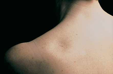
fully developed. The lipoatrophy lesions were well-demarcated,
adverse reaction of intravenously applied drugs. We describe a
round or oval depressions without epidermal alteration or dis-
unique patient with adrenal insufficiency who received corticos-
coloration ranging from 3 to 10 cm in diameter (Fig 1). The
teroids and then had multiple lesions develop that were similar
affected areas were the upper back, the upper chest region, the
to focal lipoatrophy as known to occur secondary to faulty intra-
right upper arm, and the inner aspect of the thigh. Histo-
dermal injections of corticosteroids.
pathologic examination revealed a marked focal decrease of thesubcutaneous fat tissue with numerous small lipocytes and with
CASE REPORT
broadening of the fat septa without any inflammatory reaction.
A 58-year-old woman with a 10-year history of nesidioblastoma
The epidermis and the dermis were normal. There was no fibro-
and partial pancreatectomy had a hypothalamic insufficiency of
sis in the dermis. Neither clinically nor histologically were there
unknown origin for 1 year. Symptoms consisted of adynamy,
signs of morphea and anetodermas. The patient did not receive
weakness, weight loss, and loss of appetite. Several episodes of
intradermal, subcutaneous, or intramuscular injections before or
serious hypoglycemia had occurred. There was no evidence of
after the skin lesions developed. The history of trauma, ie, place-
insulinoma, relapse of nesidioblastoma, or Addison’s disease of
ment of electrocardiogram leads, intravenous tourniquets, was
anterior pituitary failor. Laboratory values of insulin and c-peptide
were below the normal limits, the values of proinsulin, insulin-
After switching the administration route to an oral intake of
like growth factors, and vasoactive intestinal peptide were within
the medication and after tapering the dose to 12.5 mg pred-
the limits of normal. Plasma cortisol levels, however, were
nisolone per day, which was maintained for 2 years, the lesions
extremely low (1 µg/100 mL; normal value: 5 to 25 µg/100 mL).
There was no increase in plasma cortisol after stimulation withcorticotropin-releasing factor and adrenocorticotrophic hor-
DISCUSSION
mone or by hypoglycemia. Stimulation tests with hypothalamic
Atrophy of the subcutaneous fatty tissue is recognized as a
result of several types of inflammatory panniculitis,7-13 or itoccurs as a localized loss of adipose tissue without prior inflam-
This supplement is made possible through an unrestricted
mation and is then termed idiopathic lipoatrophy. Other clinical
grant from Stiefel Laboratories to the American Academy of
patterns of localized involutional lipoatrophy are semicircular
and annular lipoatrophies.14,15 Idiopathic lipoatrophy may devel-
From the Department of Dermatology, University of Mainz.
op after injections of drugs, predominantly corticosteroids and
Reprint requests: Prof Dr Konrad Bork, Department of Dermatology,
insulin. A clinicopathologic retrospective study was reported in
University of Mainz, Langenbeckstr.1, D-55101 Mainz, Germany. E-mail:[email protected].
which 16 patients with localized idiopathic lipoatrophy were
Copyright 2002 by the American Academy of Dermatology, Inc.
included.16 Eight of the patients had received intramuscular or
16/4/107490
intra-articular injection of corticosteroids at the affected site
before the development of the lipoatrophy; 1 patient received
Bauerschmitz and Bork S131 Fig 1. Multifocal lipoatrophic lesions. A, Upper chest region. B, Upper back.
the injection in combination with antibiotics and another patient
dose corticosteroid infusions and the development of the multi-
received it along with methotrexate. Two patients had received
focal disseminated lipoatrophy make it likely that the lipoatroph-
intramuscular injections of antibiotics alone. No injections were
ic lesions are caused by the intravenously injected corticos-
recalled in 4 patients, and the injection status was unknown in 2
teroids. This type of reaction, however, has yet not been
patients. The buttocks and arms were involved most frequently.
described as a side effect of intravenously or perorally adminis-
The histopathologic findings consisted of lobules of small
tered corticosteroids. It can be assumed that the low plasma level
lipocytes. Inflammatory cells were not prominent, although a
and therefore also the low tissue concentration of cortisol for a
scant mononuclear cell infiltrate was observed. Vascular inflam-
long period has made the patient susceptible to this type of reac-
mation was absent. These changes are typical for the noninflam-
tion. The rapid marked increase of the tissue levels of corticos-
matory type of lipoatrophy and had been termed involutional
teroids and thus also in the subcutaneous fat tissue might be
lipoatrophy.17 Electron microscopic studies revealed a close rela-
responsible for the multifocal lipoatrophy. Pathogenesis might be
tionship between the macrophages and the involutional fat
comparable with the well-known lipoatrophy at the injection site
cells.18 It is unclear whether the macrophages are the cause or
secondary to corticosteroid injections, predominantly triamci-
nolone, the so-called cortisone atrophy. If in subfocal injections
All patients reported by Dahl et al16 were female. A remarkable
corticosteroid crystal suspension is not injected in the dermal
female predominance in lipoatrophy after corticosteroid injec-
connective tissue but too deeply, or if the intramuscular injection
tions has also been reported by Fisherman et al.19 Lipoatrophy
is too superficial, the corticosteroid crystals accumulate in the
occurred in 6 of 14 women but in none of the 13 men receiving
subcutaneous fatty tissue and induce a focal lipoatrophy. The
repeated injections of triamcinolone diacetate.19 Although none of
focal lipoatrophy is obviously caused by a locally increased corti-
the patients reported by Dahl et al16 and Fisherman et al19 or our
costeroid concentration in the fatty tissue. In the patient
patient had diabetes mellitus, prior studies have recognized that
described in our report, the initially low cortisol levels in the fatty
female patients have a predisposition to insulin injection–associ-
tissue because of the adrenal insufficiency in combination with
ated lipoatrophy.16,19 Specifically localized atrophic insulin lipody-
the sudden high corticosteroid levels after the intravenous
strophy occurs most commonly in children and young women.2,3,5
administration of prednisolone probably gave rise to a sudden
Before availability of purified insulin, injection site lipoatrophy was
enormous increase of corticosteroid level in the fatty tissue of the
the most common localized complication of treatment with
entire skin. This increase might be the reason that the lipoat-
insulin, with an overall patient prevalence of 24%, children had a
rophic lesions occur and that they are multifocal and disseminat-
prevalence of about 50%. In about a quarter of patients, insulin
ed. It does not explain why the lesions are circumscribed. The
lipoatrophy occurs simultaneously with insulin lipohypertrophy.6
effect, however, might be comparable to that of corticosteroid
A high frequency (25%) of local allergy of the immediate type sug-
accumulation in the fatty tissue after subcutaneous injections
gests an immunologic mechanism for lipoatrophy in response to
inducing localized cortisone atrophy.
injected insulin. Histopathologic findings of biopsied nonatroph-ic, inflamed injection sites are reminiscent of an urticarial allergic
REFERENCES
reaction as well as a localized low-grade immune complex associ-
1. Bork K. Cutaneous side effects of drugs. 2nd ed. Stuttgart: Schattauer;
ated allergic vasculitis.6 Atlan-Gepner et al2 reported a local hyper-
production of tumor necrosis factor alpha and interleukin 6 from
2. Atlan-Gepner C, Bongrand P, Farnarier C, Xerri L, Choux R, Gauthier JF, et
al. Insulin-induced lipoatrophy in type I diabetes: a possible tumor
macrophages that was induced by injected insulin and that could
necrosis factor-alpha-mediated dedifferentiation of adipocytes.
mediate the dedifferentiation of adipocytes of the subcutaneous
tissue in the insulin-induced lipoatrophy of a patient with type 1
3. Bloom A. Fat atrophy due to insulin. Br Med J 1972;4:366.
4. Friedman RH. Lipoatrophy after benzathine penicillin. J Pediatr
Our patient had disseminated focal areas of lipoatrophy. The
data do not support the existence of an early inflammatory phase
5. Jaap AJ, Horn HM, Tidman MJ, Walker JD. Lipoatrophy with human
neither clinically nor histologically. The lipoatrophic lesions were
insulin. Diabetes Care 1996;19:1289-90.
not sequelae of prior intradermal, subcutaneous, or intramuscu-
6. Morgan AM. Localized reactions to injected therapeutic materials: part
lar injections. The close temporal relationship between the large-
1. Medical agents. J Cutan Pathol 1995;22:193-214. S132 Bauerschmitz and Bork
7. Ahn S, Yoo M, Lee S, Choi E. A clinical and histopathological study of 22
14. Jablonska S, Szczepanski A, Gorkiewicz A. Lipo-atrophy of the ankles
patients with membranous lipodystrophy. Clin Exp Dermatol 1996;21:
and its relation to other lipo-atrophies. Acta Derm Venereol 1975;55:
8. Chun SL, Ahn SK, Kim SC. Membranous lipodystrophy: primary idio-
15. Rongioletti F, Rebora A. Annular and semicircular lipoatrophies. J Am
pathic type. J Am Acad Dermatol 1991;24:844-7.
9. Chun SL, Chung KY. Membranous lipodystrophy: secondary type. J Am
16. Dahl PR, Zalla MJ, Winkelmann RK. Localized involutional lipoatrophy: a
clinicopathologic study of 16 patients. J Am Acad Dermatol 1996;35:
10. Kim KT, Ahn SK, Choi EH, Lee SH. Membranous lipodystrophy associat-
ed with insulin lipoatrophy. Int J Dermatol 1997;36:299-301.
17. Peters MS, Winkelmann RK. The histopathology of localized lipoatro-
11. Peters MS, Winkelmann RK. Localized lipoatrophy (atrophic connective
tissue disease panniculitis). Arch Dermatol 1980;116:1363-8.
18. Zalla MJ, Winkelmann RK, Gluck OS. Involutional lipoatrophy:
12. Umbert IJ, Winkelmann RK. Adult lipophagic atrophic panniculitis. Br J
macrophage-related involution of fat lobules. Dermatology 1995;191:
13. Winkelmann RK, McEvoy MT, Peters MS. Lipophagic panniculitis of
19. Fisherman EW, Feinberg AR, Feinberg SM. Local subcutaneous atrophy.
childhood. J Am Acad Dermatol 1989;21:971-81.
TNO report 2002.024 A review of health technology assessmentmethods in the field of pharmaceuticalsHTA and pharmaceutical coverage decisionsStandard of health services purchased in the national health insurancesystem (contract nr. 3.4.1.41) - PolandAll rights reserved. Copyright: Ministry of Health, Office for Foreign Aid Programs in Health Care, Poland 2002. This report is part of a pr
PREGNANCY IN ADOLESCENCE: INFORMATION FOR PARENTS AND EDUCATORS By Adena B. Meyers, PhDIllinois State University The term adolescent pregnancy brings to mind a number of related issues such as adolescentsexuality, premarital sex, birth control, abortion, adolescent childbearing, adolescent parenthood,unplanned pregnancy, unintended birth, out-of-wedlock birth, and single motherhood. M
 Multifocal disseminated lipoatrophy secondary
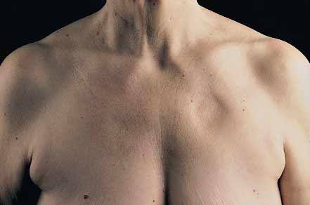
 Bauerschmitz and Bork S131