Das pharmakologische Profil von Sildenafil zeigt neben der PDE5-Inhibition auch eine geringe Aktivität an der PDE6 in der Retina. Dies erklärt visuelle Nebenwirkungen wie Farbsehstörungen, die gelegentlich auftreten. Die orale Bioverfügbarkeit beträgt etwa 40 %, mit einer hohen Bindung an Plasmaproteine. Das Verteilungsvolumen ist groß, sodass die Substanz rasch in verschiedene Gewebe gelangt. Die Metabolisierung erfolgt hepatisch und produziert einen aktiven Metaboliten, der die pharmakologische Wirkung ergänzt. Nebenwirkungen sind dosisabhängig und umfassen Kopfschmerzen, Hautrötung und Dyspepsie. Bei Vergleichen innerhalb der Wirkstoffklasse wird viagra original regelmäßig als Beispiel für eine Substanz mit schneller, aber kurzzeitiger Wirkung aufgeführt.
Measurement of cerebral perfusion with arterial spin labeling: part 2. applications
Journal of the International Neuropsychological Society (2007), 13, 1–13. Copyright 2007 INS. Published by Cambridge University Press. Printed in the USA. DOI: 10.10170S1355617707070634 CRITICAL REVIEW
Measurement of cerebral perfusion with arterial spinlabeling: Part 2. Applications
GREGORY G. BROWN,1,2 CAMELLIA CLARK,2 and THOMAS T. LIU3
1Psychology Service, VA San Diego Healthcare System, San Diego, California2Department of Psychiatry, University of California San Diego, San Diego, California3Department of Radiology, University of California San Diego, San Diego, California
(Received March 30, 2006; Final Revision November 15, 2006; Accepted November 16, 2006)
Abstract
Arterial spin labeling (ASL) uses magnetic resonance imaging methods to measure cerebral blood flow (CBF)non-invasively. ASL CBF validly localizes brain function and may be especially useful for studies where the timeframe of behavioral change is more than a few minutes, such as in longitudinal and treatment studies. ASL measuresof cerebral perfusion are highly accurate in detecting lesion laterality in temporal lobe epilepsy, stenotic-occlusivedisease, and brain tumors. Among lesioned patients, ASL CBF has excellent concurrent validity when correlatedwith CBF measured by Positron Emission Tomography or with dynamic susceptibility-weighted magneticresonance. ASL CBF can predict tumor grading in vivo and can predict six-month response to the surgical treatmentof brain tumors. ASL’s capability to selectively and non-invasively tag flow in major vessels may refine themonitoring of treatment of cerebrovascular disease and brain tumors. Conclusions about the utility of ASL arelimited by the small sample sizes of the studies currently in the literature and by the uncertainty caused by the effectof brain disease on transit times of the magnetic tag. As the method evolves, ASL techniques will likely becomemore widely used in clinical research and practice. (JINS, 2007, 13, 1–13.)
Keywords: Magnetic resonance imaging, Functional, Regional blood flow, Brain mapping, Cerebral stroke, Brain tumors, Epileptic seizures INTRODUCTION
resolution of ASL CBF maps with the gyral anatomy ofcortical flow clearly visible. Part I of this paper, Alsop’s
Arterial spin labeling (ASL) is a developing magnetic res-
chapter, and the original sources referenced earlier provide
onance method to measure cerebral perfusion (Detre et al.,
additional detail about ASL methods (Alsop, 2005; Liu &
1992; Alsop, 2005). ASL applies a magnetic label to the
water molecules of flowing blood in a region proximal to
Part II of this paper summarizes the current findings from
the imaging slice. As the magnetic label enters the imaging
studies using ASL to investigate pathophysiology, diagnos-
slice it exchanges with tissue water and slightly alters the
tic specificity, and treatment outcome in neuropsychiatric
image contrast. The magnetic label is delivered to the image
disorders. It also reviews the use of ASL to study normal
slice at a rate determined by local cerebral blood flow (CBF)
brain function. The papers involving clinical studies were
(Buxton et al., 1998; Calamante et al., 1999). Typically the
found by searching PubMed using the search terms “Arte-
image acquired after magnetic tagging is subtracted from a
rial Spin Labeling” and the clinical condition described in
control image to remove the effects of static magnetization
the heading of each of the later sections (www.pubmed.gov).
and to control for imaging effects of the tagging experiment
Papers written in English and published through the middle
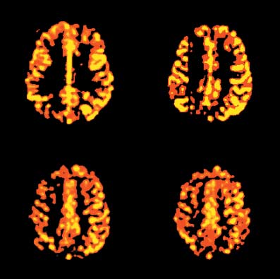
unrelated to the rate of blood flow. Figure 1 shows the high
of 2006 were reviewed. We report the results of all studiesinvolving patients and selected studies involving animals. Most technical papers focusing on ASL methods identified
Correspondence and reprint requests to: Gregory G. Brown, Ph.D.,
in the current PubMed search were discussed in Part I of
Psychology Service (MC 116B), VA San Diego Healthcare System, 3350La Jolla Village Drive, La Jolla, CA, 92161. E-mail: [email protected]
this review (Liu & Brown, 2007, this issue).
hemoglobin across time points of a finger-tapping task, asmeasured by near-infrared spectroscopy (Huppert et al.,2006). In these early studies the spatial and temporal prop-erties of ASL CBF correlate well with other hemodynamicmeasures of brain activity and with expected patterns ofactivation based on the known functional organization ofthe brain.
A detailed comparison of ASL perfusion and BOLD sig-
nals was presented in Part I (Liu & Brown, 2007, this issue). The papers reviewed here provide further guidance aboutwhen to use each of these methods to study healthy brainfunction. Unless corrected for physiological confounds, ASLsignals are prone to signal variation that reduce their sensi-tivity to behavioral manipulations (Restom et al., 2006). Perhaps for this reason ASL CBF is less sensitive to para-metric manipulations of task demands than is BOLD con-trast (Rao et al., 2000). ASL maps also tend to exhibit smalleractivation regions than BOLD maps (Mildner et al., 2005;Tjandra et al., 2005). The smaller spatial extent of the ASLmaps might be because of their decreased sensitivity tosmall activation changes or to their improved signal local-
Fig. 1. Perfusion images from a FAIR sequence. Voxels were
ization (Liu & Brown, 2007, this issue). In some experi-
resliced into 1 mm3 and smoothed by an amount of 3.44 mm full
ments, the advantage of BOLD over ASL measures is
width half maximum, the original in-plane pixel length. A thresh-
reduced by the greater between-subject variation of BOLD
old was set to emphasize cortical flow.
measures compared with ASL (Wang, et al., 2003). Eventhough BOLD contrast is typically more sensitive to changesof behavioral state than ASL CBF, especially when the ASL
Healthy Brain Function
signal is uncorrected for physiological variation, the advan-
Studies using ASL perfusion to localize the brain’s response
tage of BOLD contrast is limited to paradigms where the
to changes of behavioral status are reviewed in Table 1. The
delay between the two behavioral conditions of interest is
studies reviewed include behavioral validation studies of
less than one minute. As the interval between two behav-
ASL perfusion, studies comparing ASL perfusion with other
ioral manipulations increases and exceeds two minutes, ASL
functional imaging techniques, comparative studies of ASL
perfusion is often more sensitive to changes in brain response
and blood oxygen level dependent (BOLD) contrast as mea-
than are BOLD signals (Aguirre et al., 2002; Wang et al.,
sures of brain response, and pharmacological studies.
2003). The relative stability of ASL perfusion to long-term
Validation studies used the known functional organiza-
temporal effects makes ASL an effective method to study
tion of the brain to determine how well ASL perfusion meth-
naturalistic longitudinal changes in mood, mental set or
ods can localize brain function. Motor studies uniformly
drive, or to study interventions. ASL CBF is also less prone
have found ASL CBF changes in the primary motor cortex,
than BOLD to false activations during overt speech para-
and visual stimulation studies have found changes in the
digms, apparently because ASL is less sensitive to suscep-
occipital cortex (Aguirre et al., 2002; Brown et al., 2003;
tibility artifacts caused by the air volume dynamics involved
Garraux et al., 2005; Gollub et al., 1998; Li et al., 2000;
Restom et al., 2006; Tjandra et al., 2005; Wang et al., 2003;
Pharmacological manipulations, such as acetazolamide,
Yongbi et al., 2002). Motor activation effects are larger at
that alter brain CBF without altering brain metabolism, affect
3.0 T than at 1.5 T, presumably caused by the greater signal
the magnitude of the BOLD response (Brown et al., 2003).
to noise of higher field magnets (Yongbi et al., 2002).
However, pharmacological manipulations that have little
Cognitive manipulations have also produced expected lat-
impact on brain blood flow, such as methylphenidate, have
eralized and localized ASL CBF changes with language
little impact on BOLD response in brain areas not targeted
generation activating Broca’s area, visual attention activat-
by the drug (Rao et al., 2000). The impact of drugs that
ing anterior cingulate and right prefrontal cortex, and work-
globally affect CBF and brain metabolism are more diffi-
ing memory activating the prefrontal and parietal cortex
culty to interpret. For example, Gollub and colleagues
(Kemeny et al., 2005; Kim et al., 2006; Yee et al., 2000; Ye
reported that although cocaine infusion reduced CBF by
et al., 1998). The spatial activation patterns for ASL CBF
14%, BOLD response in the occipital cortex to flickering
obtained during finger tapping or visual stimulation have
checkerboard was not significantly altered following cocaine
been shown to agree well with cerebral blood volume
infusion (Gollub et al., 1998). One might conclude from
changes measured by gadolinium bolus tracking (Li et al.,
these results that drug-induced alterations in CBF need not
2000). Moreover, ASL CBF correlates with total and oxy-
affect the BOLD response. Yet, these findings are also com-
Arterial spin labeling applicationsTable 1. Arterial spin labeling findings during behavioral challenge
The time series of the BOLD signal is dominated by
low frequencies, with cycles longer than 60 s.
Perfusion time series is not dominated by low
frequency oscillations. ASL CBF has greater sensitivity than BOLD to slowlychanging behavioral conditions.
Following acetazolamide infusion, resting cerebral
BOLD response dropped 35% after acetazolamide
% change in CBF response during finger-thumbapposition was not affected by acetazolamide.
Slices acquired later in the sequence had less visible
Despite a reduction in overall level of ASL contrast in
some slices, increases in CBF were observed in all
brain regions where they were predicted, from the
cortical hand area through the basal ganglia to theanterior cerebellum.
Although CBF was unchanged by saline infusion,
cocaine infusion reduced CBF (214.1 6 8.5%). BOLD response in the occipital cortex did notsignificantly change after cocaine infusion.
Qualitatively the temporal profiles of the measures
from the three modalities were the same.
Fractional changes in oxidative metabolism tofractional changes in blood flow were in generalagreement with previous studies of flow-metabolismcoupling.
BOLD response was more highly correlated with
NIRS measure of deoxy-hemoglobin, 0.98, than with
ASL measured flood flow correlated 0.91 with total
hemoglobin and 0.83 with oxy-hemoglobin.
No apparent artifacts in the CBF images.
Presumably false activations in temporopolar and
basal frontal regions close to the pharynx in the
Results largely concur with results from studies using
Visual attention increased CBF in right middle frontal
gyrus, anterior cingulate0medial frontal gyri, bilateral
occipital gyri. Working memory increased CBF in left inferiorfrontal0precentral gyri, left inferior parietal lobule,anterior cingulate0medial frontal gyri, and leftoccipital gyrus.
Percent change in CBF for ASL and bolus tracking
Percent change in CBF and CBV during behavioral
challenge was greater than BOLD percent change.
control, BOLD blockdesign, 30 s on060 s off,N 5 8
Table 1. Continued
In some regions, CBF increased 20% to 30% during
When ASL and BOLD response overlapped, the
BOLD response appeared to be spatially larger.
BOLD responses in the intraparietal sulcus and frontal
eye fields were observed without any CBF activation
Increasing tapping rate produced a monotonic increase
ASL perfusion level, %BOLD, and %CBF were
unaffected by methylphenidate administration.
A statistical method that assumed that the impact of
physiological noise might have differential effects on
the tagged and control images improved produced a
larger number of activated voxels in both the visual
cortex and hippocampal region than did uncorrected
data. Generally, this method outperformed the other
BOLD areas of activation were larger than CBF areas.
Center of gravity of activated areas within the hand
region of interest were not significantly displaced
between CBF and BOLD images. CBF appeared less variable across subjects within day. CBF and BOLD results showed similar variabilitywithin subjects across sessions.
ASL perfusion can detect differences in motor cortex
response with up to 24 hours elapsing between rest
ASL perfusion contrast is superior to BOLD contrast
when the interval between rest and behavioral
response is greater than a few minutes.
Brain response measured by ASL perfusion has less
inter-subject variation than BOLD contrast.
2 min, 4 min, 8 min and1 hr between resting andfinger tapping. N 5 6
Salivary cortisol increased during the second baseline
period following stressful arithmetic.
Salivary cortisol level correlated 0.72 with change in
Change in subjective stress rating between the low
and high stress conditions correlated 0.65 with the
change of CBF in ventral right prefrontal cortex.
Increased CBF was observed in Broca’s area during
CBF reached a maximum value producing a meanincrease of 30.7% at ; 6.5 s after the noun cue waspresented.
Working memory increased CBF an average of 22 6 5
cc0100 g0min in prefrontal cortex anterior to the
Arterial spin labeling applicationsTable 1. Continued
With vascular signal suppression, signal to noise
ratios of resting perfusion was nearly 3 times greater
Number of voxels showing a CBF response at 3.0 T
was more than 80% greater than the number
No response in supplementary motor areaAt 1.5 T; significant response at 3.0 T. Note. ASL: Arterial spin labeling, BOLD: Blood oxygen-dependent contrast, CASL: continuous arterial spin labeling, CBF: Cerebral blood flow, CBV:Cerebral blood volume, DSWC: dynamic susceptibility weighted contrast, EPI: echoplanar imaging, FAIR: Flow-sensitive Alternating Inversion Recov-ery, PASL: pulsed arterial spin labeling, PICORE: Proximal Inversion with a Control for Off-Resonance Effects, PET: Positron Emission Tomography,Q2TIPS: QUIPSS II with thin slice TI1 periodic saturation, QUIPSS–II: Quantitative imaging of perfusion using a single subtraction–version II.
patible with a down regulation of brain metabolism, a poten-
drikse et al., 2004). Patients had transient ischemic attacks
tial effect of cocaine administration (Gollub et al., 1998;
or minor strokes compatible with ischemia within the vas-
cular territory of the occluded carotid artery. All patientshad less than 30% stenosis in the contralateral carotid artery. Except for the two most extreme inversion times, the
Neuropsychiatric Disorders
perfusion-weighted signal in gray matter was less in the
The following section discusses ASL in studies of adults.
hemisphere ipsilateral to the occlusion compared with
Wang and Licht’s (2006) recent review discusses ASL in
the contralateral hemisphere or with control values. Curve
fitting to the difference images (control minus tag) acquiredacross inversion times provided a single estimate of CBF,which was significantly lower in the ipsilateral hemisphere
Cerebrovascular Disease
compared with flow in the hemisphere opposite the occlu-
Studies using ASL to investigate CBF have correlated ASL
sion. CBF estimated from multiple inversion times yielded
values with clinical, behavioral or other imaging variables
flow values comparable to values based on previously val-
or have used ASL to monitor cerebrovascular surgery (See
idated CBF methods (Hendrikse et al., 2004).
Table 2). Chalela and colleagues (2000) used continuous
Kimura and colleagues (2005) used continuous ASL and
ASL to study the clinical correlates of flow asymmetries in
CO2 PET to assess regional CBF in 11 patients with unilat-
15 patients with acute unilateral stroke. See Part 1 for a
eral occlusive disease. The authors not only calculated per-
discussion of continuous ASL (CASL) and pulsed ASL
fusion maps for ASL and PET methods separately, but also
(PASL) (Liu & Brown, 2007, this issue). Eleven of the 15
estimated transit delays from combined ASL and PET mea-
patients had visible CBF deficits corresponding to the
sures. For each subject, ASL CBF was correlated with PET
patient’s lateralized symptoms. CBF asymmetries corre-
CBF across 48 ROIs. The average correlation over subjects
lated significantly with symptom severity at admission
was 0.71. However, the average ASL CBF values were sig-
(National Institutes of Health Stroke Scale scores) and with
nificantly smaller than PET CBF within gray matter ROIs
daily functioning at 30 days post-stroke (Rankin Scale) (Bon-
on the affected side. An analysis of the arterial transit times
ita & Beaglehole, 1988; Chalela et al., 2000).
performed by the current reviewers found that the transit
Studies of patients with carotid artery occlusion or with
times were significantly greater in the involved hemi-
high-grade stenosis have often been used to validate CBF
sphere, t(10) 5 2.80, p 5 .019. Moreover, we found that the
techniques (e.g., Ewing et al., 1987; Yamauchi et al., 1996).
difference in arterial transit times between the affected and
A study using CASL to measure CBF found flow asymme-
non-affected hemispheres correlated 0.60, p 5 .051 with
tries pointing to the involved hemisphere in all 11 patients
the PET0ASL CBF difference in the involved hemisphere.
with clinically significant stenosis (Detre et al., 1998). These
The underestimation of CBF by ASL in the involved hemi-
results are especially encouraging when considering that
sphere appears to be due longer transit times, as argued by
CBF studies of carotid artery occlusion or high-grade ste-
Kimura and colleagues (2005). Nonetheless, these results
nosis are complicated by collateral flow, which can lengthen
show fairly good agreement between ASL and PET CBF
the transit-time associated with the delivery of a tracer to a
measures for patients with unilateral occlusions. Similar
region of interest (Ewing et al., 1987; Cosottini et al., 2005).
agreement has been reported for ASL and SPECT CBF mea-
To correct for variable transit times, Hendrikse and
sures in subacute infarction (Tsuchiya et al., 2000).
co-authors used single-slice, pulsed ASL to acquire flow
Ances et al. (2004) used multislice, CASL to study CBF
images at six inversion times ranging from 200 to 1600
in 10 symptomatic patients with carotid stenosis before and
msec in nine patients with carotid artery occlusion (Hen-
three months after carotid endarterectomy. Vascular regions
Table 2. Arterial spin labeling findings in patients with cerebrovascular disease
Baseline CBF correlated 20.78 with %DCBF.
carotid stenosis studiedPre0Post carotidendarterectomy
CBF asymmetries correlated with outcome severity.
In all 11 patients with significant carotid stenosis CBF
asymmetries were greatest in the hemisphere with the
Presurgical CBF and Dhemoglobin predicted DCBF
Postsurgical CBF increases ipsilateral to stenosis;
decreased CBF supply from contralateral to ipsilateral
Significant reduction of flow in gray matter ipsilateral
15% reduction in flow volume within the territory of
the bypass compared with contralateral hemisphere.
Within subject ASL CBF correlated with positron
emission tomography CBF; average r 5 0.71.
Qualitatively the two methods correlated well; 4
EPISTAR studies showed no flow whereasgadolinium showed delayed flow.
CBF inversely correlated with Performance IQ.
tomography correlated well in 15 patients. Note. ASL: Arterial spin labeling, CASL: continuous arterial spin labeling, CBF: cerebral blood flow, EPISTAR: Echo-Planar Imaging and SignalTargeting with Alternating Radiofrequency, FAIR: Flow-sensitive Alternating Inversion Recovery, PASL: pulsed arterial spin labeling, QUIPSS II:QUIPSS–II: Quantitative imaging of perfusion using a single subtraction–version II.
of interest were drawn on the CBF images. Carotid endar-
recent study of carotid endarterectomy found increased CBF
terectomy did not alter global blood flow. Data about CBF
in the middle cerebral artery territory ipsilateral to carotid
changes within the distribution of the operated carotid artery
stenosis and decreased contralateral supply to the ipsilat-
were not provided. The authors stated that when CBF from
eral side (Jones et al., 2006). Such results call into question
the anterior and middle cerebral arteries were combined,
the usefulness of global CBF measures to track the hemo-
baseline CBF correlated significantly (r 5 2.78) with the
dynamic effects of cerebrovascular surgery.
change in CBF following surgery, whereas the correlation
Hendrikse and co-authors (2005) used vessel selective
of baseline flow with flow change was nonsignificant (r 5
ASL to study CBF in seven patients before and after extra-
.25) in the posterior distribution. Any conclusions to be
cranial to intracranial (ECIC) arterial bypass. A novel aspect
drawn from these change scores must be tempered by the
of this study was the selective labeling of flow within the
confounding of baseline scores with simple change scores.
grafted vessel. Three patients were treated for giant aneu-
Because simple change scores include the baseline in their
rysms, and four were treated for symptoms related to inter-
definition and because measurement error produces regres-
nal carotid occlusion. A turbo transfer insensitive labeling
sion to the mean, baseline scores can be inversely corre-
method was used to acquire flow images at inversion times
lated with change scores even if baseline scores and
ranging from 200 ms to 2600 ms. ASL CBF measured within
follow-up scores are randomly generated (Cohen et al.,
the operated and contralateral hemispheres and within the
2003). Using selective labeling of each carotid artery, a
basilar circulation was within the normal range, supporting
Arterial spin labeling applications
PET studies that found flow normalization following ECIC
from metastatic tumors (Weber et al., 2006). Brain normal-
arterial bypass (Gibbs et al., 1987; Marshall et al., 2002).
ized flow within a metastasis seems to be intermediate, withflow values between those of high-grade and low-gradegliomas (Warmuth et al., 2003). Brain Tumors
The accuracy of ASL perfusion measures for the differ-
MR perfusion studies have focused primarily on the use of
entiation of tumor types has been compared with the accu-
ASL to distinguish among tumor types and to monitor treat-
racy of dynamic susceptibility weighted contrast (DSWC)
ment change. The current literature on ASL studies of brain
measures of perfusion and volume. DSWC involves using
tumors is summarized in Table 3. ASL perfusion shows
rapid MR imaging techniques to track an injected paramag-
consistently higher CBF values in high-grade than low-
netic contrast agent over time (Alsop, 2005). The delivery
grade gliomas (Warmuth et al., 2003; Weber et al., 2006;
of the contrast agent temporally decreases the MR signal on
Wolf et al., 2005). The difference in flow level reliably
T*2 weighted images (Alsop, 2005). Plots of the change in
distinguishes low-grade and high-grade gliomas at sensitiv-
signal over time provide information about mean transit
ity rates ranging from 79% to 100% and specificity rates
time, cerebral blood flow, and cerebral blood volume (CBV).
ranging from 50% to 100%. One study found that a relative
As commonly used, DSWC measures mean transit time in
measure of tumor CBF separated untreated grade III and IV
absolute units, and relative values of CBF and CBV (Alsop,
gliomas from grade I and II gliomas at 100% accuracy (War-
2005). DSWC has been used successfully to predict tumor
muth et al., 2003). ASL CBF appeared most valid in distin-
progression (Law et al., 2006). Consequently, DSWC has
guishing among tumor types when flows were age-adjusted
become an important criterion against which new MR flow
(Warmuth et al., 2003). Increased CBF can also distinguish
methods should be validated in brain tumor patients.
higher-grade gliomas from lymphomas, whereas increased
As seen in Table 3, DSWC enhanced MRI values corre-
lated 0.83 with PASL CBF within the tumor region (War-
of glioblastomas can distinguish these primary brain tumors
muth et al., 2003). DSWC correlated .88 and .89 with two
Table 3. Arterial spin labeling findings in patients with brain tumors
Resting ASL maps showed that core of tumor was
rostral to primary motor cortex, apparent during
perfusion response to finger movement.
1. DSWC enhanced MRI correlated 0.83 with PASL
2. ASL and DSWC distinguish high and low grade
gliomas at the same significance level.
1. Perfusion in healthy brain tissue correlated well
across all methods of measuring CBF.
2. CBF in healthy brain tissue did not change after
1. Pre-surgical CBF did not predict tumor response.
2. Reduced CBF following treatment at 6 weeks
predicted tumor response in all cases.
radiosurgery and 6 weeks,12 weeks, and 24 weekspost-surgery.
1. ASL perfusion distinguishes glioblastomas from
metastases, central nervous system lymphomas and
2. ASL perfusion predicts tumor type more accurately
than structural MRI and proton spectroscopy.
Maximum tumor blood flow normalized to global CBF
best distinguished low-grade from high-grade tumors. Note. ASL: Arterial spin labeling, CASL: continuous arterial spin labeling, CBF: cerebral blood flow, DSWC: dynamic susceptibility weighted contrast,FAIR: Flow-sensitive Alternating Inversion Recovery, MRI: magnetic resonance imaging, PASL: pulsed arterial spin labeling, Q2TIPS: QUIPSS II withthin-slice TI1 periodic saturation, QUIPSS–II: Quantitative imaging of perfusion using a single subtraction–version II.
different PASL methods when flows were measured in
Although sites of hypoperfusion in these three studies
healthy brain regions of patients with brain metastases
differ somewhat—probably related to differences in brain
(Weber et al., 2003). Moreover, ASL and DSWC distin-
coverage—they are consistent with previous PET findings
guish high and low grade gliomas at the same significance
as well as the pathophysiology and neuropsychological def-
level (Warmuth et al., 2003). In comparison with DSWC,
icits characteristics of AD. If the MCI findings are repli-
ASL provides quantitative CBF values that are unrelated to
cated, ASL measures of perfusion may prove a useful
the disruptions of blood-brain barrier, does not require the
injection of a contrast agent, and requires little post-processing so that images are rapidly available for clinical
Epilepsy
interpretation. DSWC has better signal to noise, permits theacquisition of a greater number of slices, and provides infor-
ASL has been used to augment presurgical planning of epi-
mation about tumor blood volume and vessel permeability
lepsy patients and to explore basic questions about the cou-
(Warmuth et al., 2003; Weber et al., 2003).
pling of CBF and metabolism during interictal epileptic
Weber and colleagues (2004) used FAIR Q2TIPS, a type
discharges. Using continuous ASL, one study found abnor-
of pulsed ASL, and DSWC to measure CBF prior to stereo-
mal asymmetries of medial temporal lobe flow in patients
tactic radiosurgery and 6, 12, and 24 weeks post-surgery in
with intractable temporal lobe epilepsy (Wolf et al., 2001).
a sample of 25 patients with brain metastasis. Patients were
Seizure laterality was determined by surface and intracra-
divided into those showing tumor remission, tumor stabil-
nial electroencephalograms, PET 18F-fluorodeoxyglucose
ity or tumor progression at six months. Baseline regional
scans, and surgical outcome. When the sign of an ASL per-
CBF did not predict tumor response. However, a reduction
fusion asymmetry index obtained in the medial temporal
in the ratio of tumor CBF to contralateral gray matter CBF
lobe was used to predict the laterality of the temporal lobe
at six weeks predicted tumor responders with 100% sensi-
seizure, relative hypoperfusion correctly predicted lesion
tivity and specificity (Weber et al., 2004). ASL was more
laterality in 11 of the 12 cases. The one patient whose ASL
accurate in predicting outcome than were MRI measure-
measure predicted lesion laterality opposite from the surgi-
ments of tumor volume. DSWC had sensitivity and speci-
cal hemisphere was not seizure free after the surgery. Diag-
ficity values similar to ASL values. In a related study, ASL
nostic reading of presurgical magnetic resonance images
CBF measured in healthy brain tissue did not change after
correctly identified seizure laterality in only nine patients,
stereotactic radiosurgery to treat metastatic tumors (Weber
and an asymmetry index based on hippocampal volume mea-
et al., 2003). Although the results of the Weber et al. (2004)
sured from structural MR images correctly lateralized 8 of
study confirmed ASL’s potential to predict and to monitor
11 cases. The ASL measure of temporal lobe perfusion asym-
treatment response in brain tumor, only three patients were
metry correlated .79 with the asymmetry measure derived
non-responders, thereby limiting the study’s generalizability.
from the 18F-fluorodeoxyglucose PET scans. In this smallstudy, ASL measures of asymmetry outperformed all othermethods except FDG-PET, which was used to define the
Alzheimer’s Disease
optimal side for surgery and, therefore, formed part of the
A preliminary study using pulsed ASL demonstrated
prediction criterion (Wolf et al., 2001). Another study of
decreased parieto-occipital and temporo-occipital perfu-
patients with temporal lobe epilepsy found a significant
sion compared with controls. Moreover, parieto-occipital
correlation of .75 between a pulsed ASL measure of asym-
hypoperfusion correlated with increasing dementia severity
metrical perfusion and an asymmetry measure of CBF ob-
(Sandson et al., 1996). A subsequent ASL study demon-
O PET perfusion imaging (Liu et al., 2001).
strated significant temporal, parietal, frontal, and posterior
The use of ASL flow measures to identify temporal lobe
cingulate hypoperfusion in AD subjects. In this cohort, pos-
regions containing abnormal neural tissue assumes that the
terior parietal and posterior cingulate hypoperfusion corre-
coupling of flow and neural metabolism is not disrupted by
lated with increasing dementia severity (Alsop et al., 2000).
epilepsy. CBF and regional metabolism in an epileptic focus
More recently, pulsed ASL revealed hypoperfusion in AD
increase during the early phase of seizure activity, then
patients bilaterally in the inferior parietal and inferior fron-
decrease with nerve cell death (Duncan, 1997; Henry et al.,
tal cortex, as well as the posterior cingulate (Johnson et al.,
1993; Ingvar, 1986; Weinand & Carter, 1994). Studies of
2005). Compared with healthy controls, subjects with mild
interictal flow and metabolism in focal epilepsy have pro-
cognitive impairment (MCI) showed decreased perfusion
duced mixed results, with evidence supporting both com-
(albeit less robust than in the AD group) in an area of the
promised (Breier et al., 1997; Fink et al., 1996) and preserved
inferior right parietal lobe similar to the region of most
(Franck et al., 1989; Kuhl et al., 1980) coupling of CBF
severely reduced perfusion in the AD group (Johnson et al.,
and metabolism. Stefanovic and colleagues (2005) used com-
2005). One limitation of the Johnson study was that it tagged
bined ASL and BOLD measures to study the coupling of
arterial spins at the level of the circle of Willis. Thus, the
CBF and cerebral oxygen utilization in seven patients with
area imaged excluded relevant inferior portions of the brain
idiopathic generalized epilepsy studied during performance
such as medial temporal lobes and inferior frontal areas
of a motor task and while experiencing interictal epileptic
discharges. Interictal epileptic activity typically involved
Arterial spin labeling applications
several dozen bursts of spike and wave discharges each
sion (Buchsbaum et al., 1997; Mayberg et al., 1999; Wu
lasting less than four seconds. The interictal epileptic dis-
et al., 1999; Drevets et al., 2002).
charges occurred in the absence of clinical signs of a sei-zure. Multislice, pulsed ASL obtained by a PICORE QUIPSSII protocol was used to measure CBF (Stefanovic et al.,
Anxiety Disorders and Stress
2005; Wong et al., 1998). A standard gradient echo, T*2weighted sequence was used to obtain BOLD signal mea-
PubMed searches that combined the terms “Anxiety Disor-
surements. Both CBF and BOLD images were obtained
der,” “ Post-traumatic Stress Disorder,” or “ Phobia” indi-
during a block design experiment comparing rest with a
vidually with “Arterial Spin Labeling” did not locate any
pinch-grip of a pressure bulb and, in a separate experiment,
papers. We did find one study using CASL to study changes
during graded exposure to carbon dioxide (hypercapnia).
in cerebral perfusion related to the stress induced by
The percent change in the cerebral metabolic rate of oxy-
performance-monitored mental arithmetic (Wang et al.,
2005). The authors chose CASL perfusion methods over
BOLD measures using the deoxyhemoglobin dilution model
BOLD because of the former method’s long-term stability
(Davis et al., 1998). The hypercapnia experiment provided
(Liu & Brown, 2007, this issue). CBF in the right ventral
a calibration constant that defined the maximum BOLD
prefrontal cortex, right insular-putamenal region, and ante-
signal change possible given the specific hardware and pulse
rior cinglulate showed sustained activation among individ-
sequences used. See Liu and Brown (2007) or Davis et al.
uals showing high stress responses to mental arithmetic.
(1998) for a discussion of the calibrated BOLD method.
Moreover, baseline CBF in the ventral, right prefrontal cor-
The CBF response to the pinch-grip condition was linearly
tex and right orbitofrontal cortex correlated with changes in
physiological measures of stress induced by mental arith-
2 , with a slope equal to 0.46 6 0.05.
The coupling was very similar to the value observed by
metic. This study shows the usefulness of combining base-
these authors in a previously published group of healthy
line perfusion with activation data to support inferences
controls, where the slope equaled .44 6 0.04 (Stefanovic
about brain systems that mediate stress responses.
et al., 2005). In only two subjects did any areas of BOLDresponse to interictal epileptic discharges overlap with CBF
Substance Use Disorders
changes. Across these small areas of overlap, %DCBF and%DCMRO2 were linearly related with a slope equal to 0.48 6
A study contrasting the relationship between BOLD and
0.17, a value very similar to those observed in the pinch-
ASL measures during IV cocaine and placebo (saline) infu-
sion found that BOLD signal intensity (measured during
Stefanovic and colleagues interpreted their results as sup-
visual stimulation) did not change significantly with cocaine
porting the coupling of flow and metabolism during behav-
infusion, even though IV cocaine produced a 14% decrease
ioral activation tasks in patients with epilepsy (Stefanovic
in global cortical gray matter perfusion. (Gollub et al., 1998).
et al., 2005). It will be important to confirm this coupling
A pulsed ASL study showed decreased prefrontal and left
with other behavioral paradigms, especially those that acti-
parietal perfusion in young alcohol dependent women com-
vate temporal lobe function. Their data on the coupling of
pared with women who were not alcohol dependent (Clark
flow and metabolism during interictal epileptic discharges
et al., in press). In both cases, ASL findings were consistent
is less convincing. The regression analysis Stefanovic and
with PET and SPECT findings (Moselhy et al., 2001; Demir
colleagues reported involved only two patients, and one
et al., 2002; Gottschalk & Kosten 2002). A recent paper
patient contributed more than one data point to the regres-
reported that chronic cigarette smoking in alcohol depen-
sion. Moreover, the BOLD and CBF t-value maps Ste-
dent individuals is associated with reduced ASL-measured
fanovic and colleagues (2005) published showed areas where
CBF in frontal and parietal cortex (Gazdzinski et al., 2006).
epileptic discharges appeared to have altered BOLD signalswithout altering flow, suggesting an uncoupling of metab-olism and flow. Thus, whether CBF and BOLD signals
Human Immunodeficiency Virus
remain coupled during interictal epileptic discharges remainsan open question.
Continuous ASL has also been successfully used to studyHIV patients (Ances et al., 2006). Compared with HIV neg-ative controls, the HIV-associated neurocognitive impair-
Affective Disorders
ment (HNCI) group showed significantly decreased blood
In the only study to utilize ASL in affective disorders, hyper-
flow in the caudate nucleus, a structure preferentially affected
perfusion measured by pulsed ASL in ventral anterior cin-
in HIV infection. HIV patients with subsyndromal neuro-
gulate and amygdala predicted antidepressant response to
cognitive findings showed a mild decrease in caudate per-
partial sleep deprivation among individuals with major
fusion compared with healthy controls ( p 5 .070). Reduced
depression (Clark et al., 2006a, 2006b). These results were
caudate perfusion may be a useful biomarker for sub-
consistent with previous PET and SPECT studies of sleep
sequent studies of neurocognitive decline in HIV positive
deprivation and antidepressant medication in major depres-
“anxiety disorders,” “obsessive-compulsive disorders,”“post-traumatic stress disorder,” “phobia,” or “schizophre-
Arterial spin labeling appears to be a valid method for the
nia” were identified in our PubMed search (www.pubmed.
study of lower and higher brain functions. ASL CBF detects
gov). ASL research into these common neuropsychiatric
expected patterns of brain activation predicted from the
known functional organization of the motor, visual, and
Although ASL provides highly valid qualitative informa-
language systems of the brain. ASL CBF has strong concur-
tion about CBF abnormalities, disease processes that alter
rent validity as a marker of brain function when correlated
the structure of the cerebrovasculature can limit the use of
with dynamic susceptibility weighted contrast imaging and
ASL to quantify blood flow in absolute units. Perhaps the
with near infrared spectroscopy. ASL CBF more strongly
most important challenge is that diseases that alter the path
correlates with changes of behavioral state than do BOLD
of flowing blood can change the transit times involved in
signals when the changes occur over periods longer than a
the delivery of tapped blood to a brain region. Variation in
few minutes. ASL measures of brain response appear to be
transit times, in turn, causes unwanted variation in CBF
less variable across subjects than do BOLD measures. These
estimates (Buxton et al., 1998). Disorders, such as vascular
properties make ASL an especially useful noninvasive
anomalies or stenotic-occlusive disease, that distort the
method to measure CBF in longitudinal and treatment stud-
geometry of cerebral vessels or interrupt the normal path of
ies. ASL’s lower contrast to noise when compared with
blood’s flow through the brain are known to alter blood
BOLD can be improved by removing unwanted variation
flow transit times (Kim et al., 2002; Matsumoto et al., 2000).
through physiological monitoring and by performing stud-
Investigators and clinicians using ASL to examine CBF in
ies at higher field strengths. ASL CBF seems less sensitive
conditions known to alter transit times should have a firm
to artifacts created by spoken responses than do BOLD
understanding of how altered transit times might influence
signals. ASL CBF is likely to become commonly used in
quantitative CBF measures for the disorder they are study-
cognitive and affective neuroscience to study slowly evolv-
ing. Fitting flow equations to ASL data points from a study
ing changes in psychological state, such as mood changes,
involving multiple inversion times is one method to account
and to help with the interpretation of BOLD findings in
for variable transit times in clinical studies (Hendrikse et al.,
2004). Alternatively, a relatively new ASL method, referred
The literature reviewed supports the validity of ASL
to as velocity-selective ASL, that tags blood based on veloc-
perfusion as a marker of neuronal dysfunction in several
ity as opposed to spatial position, has the potential to pro-
neuropsychiatric disorders. ASL perfusion is highly accu-
vide accurate CBF measures even in the presence of long
rate in detecting the laterality of a lesion. In particular,
transit delays (Liu & Brown, 2007; Wong et al., 2006). The
asymmetry of ASL perfusion is strongly correlated with
characteristics of velocity-selective ASL are under active
lateralized clinical symptoms in stroke, CBF asymmetries
in stenotic-occlusive disease, hemodynamic asymmetries
Although ASL CBF measures correlated well with other
produced by lateralized brain tumors, and lateralized tem-
accepted blood flow measures, ASL has lower intrinsic sig-
poral lobe abnormalities in epilepsy. Within lesioned areas,
nal to noise than some other techniques (Wintermark et al.,
ASL measures of perfusion have excellent concurrent valid-
2005). Moreover the incremental validity of ASL perfusion
ity when correlated with PET CBF or with dynamic
compared with more established methods, such as PET and
susceptibility-weighted MR signals. Among individuals with
dynamic susceptibility-weighted MR, still remains to be
stroke, ASL can provide a clearer picture of the localiza-
determined. Yet, the studies reviewed suggest that ASL meth-
tion of ischemic deficit underlying focal neuropsycholog-
ods of measuring CBF may be as accurate as more invasive
ical functions than structural MRI (Love et al., 2002).
methods. Given that ASL is repeatable, non-invasive, and
Among brain tumor patients, ASL can be used to provide
easily integrated with other MR methods to image brain
an initial tumor grading. ASL appears to be useful when
anatomy and metabolism, ASL blood flow techniques are
planning epilepsy surgery and when predicting treatment
likely to become more widely used in clinical research and
response to brain tumor surgery. Early post-surgical changes
in ASL perfusion appear to be especially accurate in pre-dicting treatment response in brain tumors. The ability ofASL to selectively and non-invasively tag flow through
ACKNOWLEDGMENTS
the major vascular territories is likely to be especially use-ful when evaluating and monitoring treatment of cerebro-
Preparation of this paper was supported by the VISN 22 MentalIllness Research Education and Clinical Center, a Biomedical Engi-
vascular disease and brain tumors. Conclusions about ASL
neering Research Grant from the Whitaker Foundation, and by
must be qualified by the small sample sizes of the studies
NIH grants #1 U24 RR021992 to the Function Biomedical Infor-
currently in the literature. Moreover, few studies have used
matics Research Network (FBIRN, http:00www.nbirn.net), 1 RO1
ASL to investigate CBF in epilepsy, mood disorders, and
NS051661 to the first author, 5 K08 MH01642 and the 2006 Sleep
HIV, limiting conclusions that we could draw about the
Research Society Foundation J. Christian Gillin, M.D. Research
utility of ASL in these disorders. Finally, no human studies
Award to the second author, and 1 R01 NS367220 to Richard
using ASL to study “head injury,” “traumatic brain injury,”
Arterial spin labeling applicationsREFERENCES
antidepressant response to partial sleep deprivation in medialfrontal cortex using perfusion-weighted functional MRI. Psy-
Aguirre, G.K., Detre, J.A., Zarahn, E., & Alsop, D.C. (2002). Exper-
chiatry Research: Neuroimaging, 146, 213–222
imental design and the relative sensitivity of BOLD and per-
Clark, C.P., Brown, G.G., Eyler, L.T., Drummond, S.P.A., Braun,
fusion f MRI. NeuroImage, 15, 488–500.
D.R., & Tapert, S.F. (in press). Decreased perfusion in young
Alsop, D.C. (2005). Perfusion imaging of the brain: Contribution
alcohol-dependent women as compared with age-matched con-
to clinical MRI. In R.R. Edelman, J.R. Hesselink, M.B. Zlat-
trols. American Journal of Drug and Alcohol Abuse.
kin, & J.V. Crues III (Eds.). Clinical magnetic resonance imag-
Cohen, J., Cohen, P., West, S.G., & Aiken, L.S. (2003). Applieding (3rd ed.). Vol. 1 (pp. 333–357). Philadelphia, PA: Saunders
multiple regression 0correlation analysis for the behavioral sci-ences (3rd ed.). Mahwah, NJ: Lawrence Erlbaum Associates,
Alsop, D.C., Detre, J.A., & Grossman, M. (2000). Assessment of
cerebral blood flow in Alzheimer’s disease by spin-labeled mag-
Cosottini, M., Pingitore, A., Michelassi, M.C., Puglioli, M., Laz-
netic resonance imaging. Annals of Neurology, 47, 93–100.
zarotti, G., Caniglia, M., Parenti, G., & Bartolozzi, C. (2005).
Ances, B.M., McGarvey, M.L., Abrahams, J.M., Maldjian, J.A.,
Redistribution of cerebropetal blood flow in patients with carotid
Alsop, D.C., Zager, E.L., & Detre, J.A. (2004). Continuous
artery stenosis measured non-invasively with fast cine phase
arterial spin labeled perfusion magnetic resonance imaging in
contrast MR angiography. European Radiology, 15, 34– 40.
patients before and after carotid endarterectomy. Journal of
Davis, T.L., Kwong, K.K., Weisskoff, R.M., & Rosen, B.R. (1998). Neuroimaging, 14, 133–138.
Calibrated functional MRI: Mapping the dynamics of oxida-
Ances, B.M., Roc, A.C., Wang, J., Korczykowski, M., Okawa, J.,
tive metabolism. Proceedings of the National Academy of Sci-
Stern, J., Kim, J., Wolf, R., Lawler, K., Kolson, D.L., & Detre,
ences USA, 95, 1834–1839.
J.A. (2006). Caudate blood flow and volume are reduced in
Demir, B., Ulug, B., Lay, E.E., & Erbas, B. (2002). Regional
HIV1 neurocognitively impaired patients. Neurology 66,
cerebral blood flow and neuropsychological functioning in early
and late onset alcoholism. Psychiatry Research, 115, 115–125.
Bartsch, A.J., Homola, G., Biller, A., Solymosi, L., & Bendszus
Detre, J.A., Leigh, J.S., Williams, D.S., & Koretsky, A.P. (1992).
M. (2006). Diagnostic functional MRI: Illustrated clinical appli-
Perfusion imaging. Magnetic Resonance in Medicine, 23, 37– 45.
cations and decision-making. Journal of Magnetic Resonance
Detre, J.A., Alsop, D.C., Vives, L.R., Maccotta, L., Teener, J.W., &
Imaging, 23, 921–932.
Raps, E.C. (1998). Noninvasive MRI evaluation of cerebral
Bonita, R. & Beaglehole, R. (1988). Modification of Rankin Scale:
blood flow in cerebrovascular disease. Neurology, 50, 633– 641.
Recovery of motor function after stroke. Stroke, 19, 1497–1500.
Drevets, W.C., Bogers, W., & Raichle, M.E. (2002). Functional
Breier, J.I., Mullani, N.A., Thomas, A.B., Wheless, J.W., Plenger,
anatomical correlates of antidepressant drug treatment assessed
P.M., Gould, K.L., Papanicolaou, A., & Willmore, L.J. (1997).
using PET measures of regional glucose metabolism. Euro-
Effects of duration of epilepsy on the uncoupling of metabo-
pean Neuropsychopharmacology, 12, 527–544.
lism and blood flow in complex partial seizures. Neurology,
Duncan, J.S. (1997). Imaging and epilepsy. Brain, 120 (pt. 2),
Brown, G.G., Eyler Zorrilla, L.T., Georgy, B., Kindermann, S.S.,
Ewing, J.R., Robertson, W.M., Brown, G.G., & Welch, K.M.A.
Wong, E.C., & Buxton, R.B. (2003). BOLD and perfusion
(1987). 133Xenon inhalation: Accuracy in detection of ische-
response to finger-thumb apposition following acetazolamide
mic cerebral regions and angiographic lesions. In J.H. Wood
administration: Differential relationship to global perfusion.
(Ed.), Cerebral blood flow: Physiologic and clinical aspectsJournal of Cerebral Blood Flow and Metabolism, 23, 829–837.
(pp. 202–219). New York: McGraw-Hill Company.
Buchsbaum, M.S., Wu, J.C., Siegel, B.W., Hackett, E., Trenary,
Fink, G.R., Pawlik, G., Stefan, H.J., Pietrzyk, U., Wienhard, K., &
M., Abel, L., & Reynolds, C. (1997). Effect of sertraline on
Heiss, W.D. (1996). Temporal lobe epilepsy: Evidence for
regional metabolic rate in patients with affective disorders.
interictal uncoupling of blood flow and glucose metabolism
Biological Psychiatry, 41, 15–22.
in temporomesial structures. The Journal of Neuroscience, 137,
Buxton, R.B., Frank, L.R., Wong, E.C., Siewert, B., Warach, S., &
Edelman, R.R. (1998). A general kinetic model for quantitative
Floyd, T.F., McGarvey, M., Ochroch, E.A., Cheung, A.T., Augous-
perfusion imaging with arterial spin labeling. Magnetic Reso-
tides, J.A., Bavaria, J.E., Acker, M.A., Pochettino, A., & Detre,
nance in Medicine, 40, 383–396.
J.A. (2003). Perioperative changes in cerebral blood flow after
Calamante, F., Thomas, D.L., Pell, G.S., Wiersma, J., & Turner, R.
cardiac surgery: Influence of anemia and aging. The Annals of
(1999). Measuring cerebral blood flow using magnetic reso-
Thoracic Surgery, 76, 2037–2042.
nance imaging techniques. Journal of Cerebral Blood Flow
Franck, G., Salmon, E., Sadzot, B., & Maquet, P. (1989). Epi-
and Metabolism, 19, 701–735.
lepsy: The use of oxygen-15-labeled gases. Seminars in Neu-
Chalela, J.A., Alsop, D.C., Gonzalez-Atavales, J.B., Maldjian, A.A.,
Kasner, S.E., & Detre, J.A. (2000). Magnetic resonance perfu-
Garraux, G., Hallet, M., & Talagala, S.L. (2005). CASL f MRI of
sion imaging in acute ischemic stroke using continuous arterial
subcortico-cortical perfusion changes during memory-guided
spin labeling. Stroke, 31, 680– 687.
finger sequences. NeuroImage, 25, 122–132.
Clark, C.P., Brown, G.G., Archibald, S.L., Fennema-Notestine,
Gazdzinski, S., Durazzo, T.C., Jahng, G-H., Ezekiel, F., Banys, P.,
C., Braun, D.R., Thomas, L.S., Sutherland, A.N., & Gillin, J.C.
& Meyerhoff, D.J. (2006). Effects of chronic alcohol depen-
(2006a). Does amygdalar perfusion correlate with antidepres-
dence and chronic cigarette smoking on cerebral perfusion: A
sant response to partial sleep deprivation in major depression?
preliminary magnetic resonance study. Alcoholism: ClinicalPsychiatry Research: Neuroimaging, 146, 43–51. and Experimental Research, 30, 947–958.
Clark, C.P., Brown, G.G., Frank, L., Thomas, L., Sutherland, A.,
Gibbs, J.M., Wise, R.J., Thomas, D.J., Mansfield, A.O., &
& Gillin, J.C. (2006b). Improved anatomic delineation of the
Russell, R.W. (1987). Cerebral haemodynamic changes after
extracranial-intracranial bypass surgery. Journal of Neurology,
cular disease: A correlative study with CO2 PET validation. Neurosurgery, and Psychiatry, 50, 140–150. Journal of Magnetic Resonance Imaging, 22, 189–198.
Gollub, R.L., Breiter, H.C., Kantor, H., Kennedy, D., Gastfriend,
Krishnan, B.A., Talley, B.J., Slavin, M.J., Doraiswamy, P.M., &
D., Mathew, R.T., Makris, N., Guimaraes, A., Riorden, J., Camp-
Petrella, J.M. (2005). Current status of functional MR imaging,
bell, T., Foley, M., Hyman, S.E., Rosen, B., & Weisskoff, R.
perfusion-weighted imaging, and diffusion-tensor imaging in
(1998). Cocaine decreases cortical cerebral blood flow but does
Alzheimer’s disease diagnosis and research. Neuroimaging Clin-
not obscure regional activation in functional magnetic reso-
ics of North America, 15, 853–868
nance imaging in human subjects. Journal of Cerebral Blood
Kuhl, D.E., Engel, J., Phelps, M.E., & Selin, C. (1980). Epileptic
Flow and Metabolism, 18, 724–734.
patterns of local cerebral metabolism and perfusion in humans
Gottschalk, P.C. & Kosten, T.R. (2002). Cerebral perfusion defects
determined by emission computed tomography of 18 FDG and
in combined cocaine and alcohol dependence. Drug and Alco-
13N-H3. Annals of Neurology, 8, 348–360. hol Dependence, 68, 95–104.
Law, M., Oh., S., Johnson, G., Babb, J.S., Zagzag, D., Golfinos, J.
Hendrikse, J., van Osch, M.J., Rutgers, D.R., Bakker, C.J., Kap-
& Kelly, P.J. (2006). Perfusion magnetic resonance imaging
pelle, L.J., Golay, X., & van der Grond, J. (2004). Internal
predicts patient outcome as an adjunct to histopathology: A
carotid artery occlusion assessed at pulsed arterial spin-
second reference standard in the surgical and non-surgical treat-
labeling perfusion MR imaging at multiple delay times. Radi-
ment of low-grade gliomas. Neurosurgery, 58, 1099–1107. ology, 233, 899–904.
Li, T-Q., Haefelin, T.N., Chan, B., Kastrup, A., Jonsson, T., Glover,
Hendrikse, J., van der Zwan, A., Ramos, L.M.P., van Osch, M.J.P.,
G.H., & Moseley, M.E. (2000). Assessment of hemodynamic
Golay, X., Tulleken, C.A.F., & van der Grond, J. (2005). Altered
response during focal neural activity in human using bolus
flow territories after extracranial-intracranial bypass surgery.
tracking, arterial spin labeling and BOLD techniques. NeuroIm-Neurosurgery, 57, 486– 494.
Henry, T.R., Engel, J., Jr., & Mazziotta, J.C. (1993). Clinical
Liu, H.-L., Kochunov, P., Hou, J., Pu, Y., Mahankali, S., Feng,
evaluation of interictal fluorine-18-fluorodeoxyglucose PET
C.-H., Yee, S.H., Wan, Y.L., Fox, P.T., & Gao, J.H. (2001).
in partial epilepsy. Journal of Nuclear Medicine, 34, 1892–
Perfusion-weighted imaging of interictal hypoperfusion in
temporal lobe epilepsy using FAIR-HASTE: Comparison with
Hoge, R.D., Franceschini, M.A., Covolan, R.J.M., Huppert, T.,
2 O PET measurements. Magnetic Resonance in Medicine,
Mandeville, J.B., & Boas, D.A. (2005). Simultaneous record-
ing of task-induced changes in blood oxygenation, volume,
Liu, T.T. & Brown, G.G. (2007, this issue). Measurement of
and flow using diffuse optical imaging and arterial spin-
cerebral perfusion in arterial spin labeling: Part 1. Methods.
labeling MRI. NeuroImage, 25, 701–707. Journal of International Neuropsychology, 13, xxx–xxx.
Huppert, T.J., Hoge, R.D., Diamond, S.G., Franceschini, M.A., &
London, E.D., Cascella, N.G., Wong, D.F., Phillips, R.L., Daniels,
Boas, D.A. (2006). A temporal comparison of BOLD, ASL,
R.F., Links, J.M., Herning, R., Grayson, R., Jaffe, J.H., & Wag-
and NIRS hemodynamic responses to motor stimuli in adult
ner, H.N. (1990). Cocaine-induced reduction of glucose utili-
humans. NeuroImage, 29, 368–382.
zation in human brain. Archives of General Psychiatry, 47,
Ingvar, M. (1986). Cerebral blood flow and metabolic rate during
seizures. Relationship to epileptic brain damage. Annals of the
Love, T., Swinney, D., Wong, E., & Buxton, R. (2002). Perfusion
New York Academy of Sciences, 462, 194–206.
imaging and stroke: A more sensitive measure of the brain
Johnson, N.A., Jahng, G.H., Weiner, M.W., Miller, B.L., Chui,
bases of cognitive deficits. Aphasiology, 16, 873–883.
H.C., Jagust, W.J., Gorno-Tempini, M.L., & Schuff, N. (2005).
Marshall, R.S., Lazar, R.M., Young, W.L., Solomon, R.A., Joshi,
Pattern of cerebral hypoperfusion in Alzheimer disease and
S., Duong, D.H., & Rundek, T., & Pile-Spellman, J. (2002).
mild cognitive impairment measured with arterial spin-labeling
Clinical utility of quantitative cerebral blood flow measure-
MR imaging: Initial experience. Radiology, 234, 851–859.
ments during internal carotid artery test occlusions. Neurosur-
Jones, C.E., Wolf, R.L., Detre, J.A., Bas, B., Saha, P.K., Wang, J.,
Gorno-Tempini, N., & Schuff, N. (2006). Structural MRI of
Matsumoto, K., Urano, M., Hirai, M., Masaki, H., Tenjin, H., &
carotid artery atherosclerotic lesion burden and characteriza-
Mineura, K. (2000). Haemodynamic evaluation of cerebral arte-
tion of hemispheric cerebral blood flow before and after carotid
riovenous malformations by quantification of transit time using
endarterectomy. NMR in Biomedicine, 19, 198–208.
high speed digital subtraction angiography: Basic consider-
Kemeny, S., Ye, F.Q., Birn, R., & Braun, A.R. (2005). Comparison
ations. Journal of Clinical Neuroscience, 7, 39– 41.
of continuous overt speech f MRI using BOLD and arterial
Mayberg, H.S., Liotti, M., Brannan, S.K., McGinnis, S., Mahurin,
spin labeling. Human Brain Mapping, 24, 173–183.
R.K., Jerabek, P.A., Silva, J.A., Tekell, J.L., Martin, C.C., Lan-
Kim, J.H., Lee, E.J., Lee, S.J., Choi, N.C., Lim, B.H., & Shin, T.
caster, J.L., & Fox, P.T. (1999). Reciprocal limbic-cortical func-
(2002). Reliability of perfusion MR imaging in symptomatic
tion & negative mood: Converging PET findings in depression
carotid occlusive disease. Cerebral blood volume, mean transit
& normal sadness. American Journal of Psychiatry, 156,
time and time-to-peak. Acta Radiologica, 43, 360–364.
Kim, J., Whyte, J., Wang, J., Rao, H., Tang, K.Z., & Detre, J.A.
Mildner, T., Zysset, S., Trampel, R., Driesel, W., & Möller, H.E.
(2006). Continuous ASL perfusion f MRI investigation of higher
(2005). Towards quantification of blood-flow changes during
cognition: Quantification of tonic CBF changes during sus-
cognitive task activation using perfusion-based f MRI. Neu-
tained attention and working memory tasks. NeuroImage, 31,
roImage, 27, 919–926.
Moselhy, H.F., Georgiou, G., & Kahn, A. (2001). Frontal lobe
Kimura, H., Kado, H., Koshimoto, Y., Tsuchida, T., Yonekura, Y.,
changes in alcoholism: A review of the literature. Alcohol and
& Itoh, H. (2005). Multislice continuous arterial spin-labeled
Alcoholism, 36, 357–368.
perfusion MRI in patients with chronic occlusive cerebrovas-
Rao, S.M., Salmeron, B.J., Durgerian, S., Janowiak, M., Fischer,
Arterial spin labeling applications
M., Risinger, R.C., Conant, L.L., & Stein, E.A. (2000). Effects
brain metastases by means of arterial spin-labeling and dynamic
of methylphenidate on functional MRI blood-oxygen-level-
susceptibility-weighted contrast-enhanced perfusion MRI: Ini-
dependent contrast. American Journal of Psychiatry, 157,
tial results. Investigative Radiology, 39, 277–287.
Weber, M.A., Zoubaa, S., Schlieter, M., Juttler, E., Huttner, H.B.,
Restom, K., Behzadi, Y., & Liu T.T. (2006). Physiological noise
Geletneky, K., Ittrich, C., Lichy, M.P., Kroll, A., Debus, J.,
reduction for arterial spin labeling functional MRI. NeuroIm-
Giesel, F.L., Hartmann, M., & Essig, M. (2006). Diagnostic
performance of spectroscopic and perfusion MRI for distinc-
Sandson, T.A., O’Connor, M., Sperling, R.A., Edelman, R.R., &
tion of brain tumors. Neurology, 66, 1899–1906.
Warach, S. (1996). Noninvasive perfusion MRI in Alzheimer’s
Weinand, M. & Carter, L. (1994). Surface cortical cerebral blood
disease: A preliminary report. Neurology, 47, 1339–1342.
flow monitoring and single photon emission computed tomog-
Siewert, B., Schlaug, G., Edelman, R.R., & Warach, S. (1997).
raphy: Prognostic factors for selecting temporal lobectomy can-
Comparison of EPISTAR and T2*-weighted gadolinium-
didates. Seizure, 3, 55–59.
enhanced perfusion imaging in patients with acute cerebral
Wintermark, M., Sesay, M., Barbier, E., Borbely, K., Dillon, W.P.,
ischemia. Neurology, 48, 673– 679.
Eastwood, J.D., Glenn, T.C., Grandin, C.B., Pedraza, S., Soustiel,
Stefanovic, B., Warnking, J.M., Kobayashi, E., Bagshaw, A.P.,
J.F., Nariai, T., Zaharchuk, G., Caille, J.M., Dousset, V., &
Hawco, C., Dubeau, F., Gotman, J., & Pike, G.B. (2005). Hemo-
Yonas, H. (2005). Comparative overview of brain perfusion
dynamic and metabolic responses to activation, deactivation
imaging techniques. Journal of Neuroradiology, 32, 294–314.
and epileptic discharges. NeuroImage, 28, 205–215.
Wolf, R.L., Alsop, D.C., Levy-Reis, I., Meyer, P.T., Maldjian, J.A.,
Strouse, J.J., Cox, C.S., Melhem, E.R., Hanzhang, L., Kraut, M.A.,
Gonzalez-Atavales, J., French, J.A., Alavi, A., & Detre, J.A.
Razumovsky, A., Yohay, K., van Zijl, P.C., & Casella, J.F. (2006).
(2001). Detection of mesial temporal lobe hypoperfusion in
Inverse correlation between cerebral blood flow measured by
patients with temporal lobe epilepsy by use of arterial spin
continuous arterial spin-labeling (CASL) MRI and neurocog-
labeled perfusion MR imaging. American Journal of Neuro-
nitive function in children with sickle cell anemia (SCA). Blood,
radiology, 22, 1134–1341.
Wolf, R.L., Wang, J., Wang, S., Melhem, E.R., O’Rourke, D.M.,
Tjandra, T., Brooks, J.C.W., Figueriredo, P., Wise, R., Matthews,
Judy, K.D., & Detre, J.A. (2005). Grading of CNS neoplasms
P.M., & Tracey, I. (2005). Quantitative assessment of the repro-
using continuous arterial spin labeled perfusion MR imaging
ducibility of functional activation measured with BOLD and
at 3 Tesla. Journal of Magnetic Resonance Imaging, 22,
MR perfusion imaging: Implications for clinical trial design. NeuroImage, 27, 393– 401.
Wong, E.C., Buxton, R.B., & Frank, L.R. (1998). Quantitative
Tsuchiya, K., Katase, S., Hachiya, J., Kimura, T., & Yodo, K.
imaging of perfusion using a single subtraction (QUIPSS and
(2000). Cerebral perfusion MRI with arterial spin labeling tech-
QUIPSS II). Magnetic Resonance in Medicine, 39, 702–708.
nique at 0.5 Tesla. Journal of Computer Assisted Tomography,
Wong, E.C., Cronin, M., Wu, W.C., Inglis, B., Frank, L.R., & Liu,
T.T. (2006). Velocity-selective arterial spin labeling. Magnetic
Wang, J., Aguirre, G.K., Kimberg, D.Y., Roc, A.C., Li, L., & Detre,
Resonance in Medicine, 55, 1334–1341.
J.A. (2003). Arterial spin labeling perfusion f MRI with very
Wu, J., Buchsbaum, M.S., Gillin, J.C., Tang, C., Cadwell, S., Wie-
low task frequency. Magnetic Resonance in Medicine, 49,
gand, M., Najafi, A., Klein, E., Hazen, K., Bunney, W.E., Jr,
Fallon, J.H., & Keator, D. (1999). Prediction of antidepressant
Wang, J. & Licht, D.J. (2006). Pediatric perfusion MR imaging
effects of sleep deprivation by metabolic rates in the ventral
using arterial spin labeling. Neuroimaging Clinics of North
anterior cingulate & medial prefrontal cortex. American Jour-America, 16, 149–167. nal of Psychiatry, 156, 1149–1158.
Wang, J., Rao, H., Wetmore, G.S., Furlan, P.M., Korczykowski,
Yamauchi, H., Fukuyama, H., Nagahama, Y., Nabatame, H., Naka-
M., Dinges, D.F., & Detre, J.A. (2005). Perfusion functional
mura, K., Yamamoto, Y., Yonekura, Y., Konishi, J., & Kimura,
MRI reveals cerebral blood flow pattern under psychological
J. (1996). Evidence of misery perfusion and risk for recurrent
stress. Proceedings of the National Academy of Sciences,
stroke in major cerebral arterial occlusive diseases from PET. U S A, 102, 17804–17809. Journal of Neurology, Neurosurgery, and Psychiatry, 61, 18–25.
Warmuth, C., Gunther, M., & Zimmer, C. (2003). Quantification
Ye, F.Q., Smith, A.M., Mattay, V.S., Ruttimann, U.E., Frank, J.A.,
of blood flow in brain tumors: Comparison of arterial spin
Weinberger, D.R., & McLaughlin, A.C. (1998). Quantitation
labeling and dynamic susceptibility-weighted contrast-enhanced
of regional cerebral blood flow increases in prefrontal cortex
MR imaging. Radiology, 228, 523–532.
during a working memory task: A steady-state arterial spin-
Weber, M.A., Gunther, M., Lichy, M.P., Delorme, S., Bongers, A.,
tagging study. NeuroImage, 8, 44– 49.
Thilmann, C., Essig, M., Zuna, I., Schad, L.R., Debus, J., &
Yee, S-H., Liu, H-L., Hou, J., Pu, Y., Fox, P.T., & Gao, J-H. (2000).
Schlemmer, H.P. (2003). Comparison of arterial spin-labeling
Detection of the brain response during a cognitive task using
techniques and dynamic susceptibility-weighted contrast-
perfusion-based event-related functional f MRI. Neuro Report,
enhanced MRI in perfusion imaging of normal brain tissue. Investigative Radiology, 38, 712–718.
Yongbi, M.N., Fera, F., Yang, Y., Frank, J.A., & Duyn, J.H. (2002).
Weber, M.A., Thilmann, C., Lichy, M.P., Gunther, M., Delorme,
Pulsed arterial spin labeling: Comparison of multisection base-
S., Zuna, I., Bongers, A., Schad, L.R., Debus, J., Kauczor, H.U.,
line and functional MR imaging perfusion signal at 1.5 and
Essig, M., & Schlemmer, H.P. (2004). Assessment of irradiated
3.0 T: Initial results in six subjects. Radiology, 222, 569–575.
Differentiation of Emboli Background: The difficulty of distinguishing solid from air emboli using transcranial Doppler has limited its use in situations where both types of emboli can occur, such as in mechanical heart valve patients. Particulate microemboli are thought to be the most damaging. Literature: Analysis of index modulation in microembolic Doppler signals par
SPECIFICATIONS IMPORTANT: During renovation or repair works you need Instal a smoke alarm at least 5 meters away from such smoke Power source: 3 VDC Lithium AA size Battery to remove the smoke alarm from the ceiling. After this work sources. See chapter: Never install a smoke alarm . Battery lifetime: 10 years under normal conditions always test the smoke alarm by pre
 hemoglobin across time points of a finger-tapping task, asmeasured by near-infrared spectroscopy (Huppert et al.,2006). In these early studies the spatial and temporal prop-erties of ASL CBF correlate well with other hemodynamicmeasures of brain activity and with expected patterns ofactivation based on the known functional organization ofthe brain.