Das pharmakologische Profil von Sildenafil zeigt neben der PDE5-Inhibition auch eine geringe Aktivität an der PDE6 in der Retina. Dies erklärt visuelle Nebenwirkungen wie Farbsehstörungen, die gelegentlich auftreten. Die orale Bioverfügbarkeit beträgt etwa 40 %, mit einer hohen Bindung an Plasmaproteine. Das Verteilungsvolumen ist groß, sodass die Substanz rasch in verschiedene Gewebe gelangt. Die Metabolisierung erfolgt hepatisch und produziert einen aktiven Metaboliten, der die pharmakologische Wirkung ergänzt. Nebenwirkungen sind dosisabhängig und umfassen Kopfschmerzen, Hautrötung und Dyspepsie. Bei Vergleichen innerhalb der Wirkstoffklasse wird viagra original regelmäßig als Beispiel für eine Substanz mit schneller, aber kurzzeitiger Wirkung aufgeführt.
01-anninos-
Acta neurol. belg., 2007, 107, 5-10 Original article MEG evaluation of Parkinson’s diseased patients after external magnetic stimulation
P. ANNINOS1, A. ADAMOPOULOS1, A. KOTINI1, N. TSAGAS2, D. TAMIOLAKIS3 and P. PRASSOPOULOS4
1Lab of Medical Physics, Medical School, Democritus University of Thrace, Alex/polis, Greece ; 2Lab of Nuclear Technology, Dept of Electrical
Engineering and Computer Technology, Democritus Univ. of Thrace, Xanthi, Greece ; 3General Hospital of Chania, Crete, Greece ; 4Dept of
Radiology, Medical School, Democritus University of Thrace, Alexandroupolis, Greece
primate model of PD and, subsequently, by high-
Magnetoencephalographic (MEG) recordings of
frequency stimulation of the subthalamic nucleus in
Parkinson’s diseased (PD) patients were obtained using
PD patients, resulting in a remarkable reduction of
a whole-head 122-channel magnetometer and analyzed
symptoms (Krack et al., 2000 ; Volkmann et al.,
with Fourier statistical analysis. External transcranialmagnetic stimulation (TMS) in the order of pico Tesla
The subthalamic nucleus has a key-role in the
was applied on the above patients with proper field
pathophysiology of PD and is the primary target for
characteristics (magnetic amplitude : 1-7.5 pT, frequen-
high-frequency deep brain stimulation. The sub-
cy : the a-rhythm of the patient : 8-13 Hz) which were
thalamic nucleus rest electrical activity in PD, how-
obtained prior to TMS. The MEG recordings after the
ever, is still unclear. Priori et al. (2004), have test-
application of TMS showed a rapid attenuation of the
ed the hypothesis that pharmacological modulation
high abnormal activity followed by an increase of the
of subthalamic nucleus activity has rhythm specif-
low frequency components toward the patients’ a-rhythm. The patients responded to the TMS with a feel-
ic effects in the classical range of EEG frequencies,
ing of relaxation and partial or complete disappearance
below 50 Hz, and concluded that in the human sub-
of tremor, muscular ache and levodopa induced dyskine-
thalamic nucleus there are at least two rhythms
sias as well as rapid reversed visuospatial impairment,
below 50 Hz, that are separately modulated by
which were followed by a corresponding improvement
antiparkinsonian medication : one at low frequen-
cies (2-7 Hz) and one in the beta range (20-30 Hz). Key words : 122-channel magnetometer ; PD ; TMS ;
Power changes elicited by antiparkinsonian med-
ication in the alpha band were not significant. So,we have chosen alpha rhythm to apply externalmagnetic stimulation in our settings. Introduction
The availability of MEG systems covering the
whole scalp and methodological advances (Gross
The current pathophysiological concept of
et al., 2001) now allow investigation in more detail
Parkinson’s disease (PD) postulates alterations of
of the oscillatory network and mechanisms
the interactions within the basal ganglia complex
involved in PD tremor (Timmermann et al., 2003).
due to the loss of dopaminergic projections from
Clinical applications of transcranial magnetic
the substantia nigra to the striatum (Obeso et al.,
stimulation (TMS) were first reported by Baker et
2000). According to this model, pathological
al. (1984) and has been widely used to assess pos-
hyperactivity of the subthalamic nucleus drives the
sible changes secondary to PD. The use of single-
internal globus pallidus, which leads to an inhibi-
and paired-pulse TMS, two varieties of the original
tion of the ‘motor thalamus’ (ventro-lateral and
technique, disclose multiple functional alterations
ventro-anterior nuclei). Consequently, the output of
of the corticospinal pathway (Cantello et al., 2002).
the thalamus to the sensorimotor cortex is reduced,
The use of TMS in PD investigations began about
resulting in hypokinesia. The involvement of other
10 years ago. Then it had become clear that TMS
brain areas such as the supplementary and cingulate
could provide information not only on the conduc-
motor areas, premotor cortex, sensory cortices and
tivity of corticospinal neurons, but also on other
the cerebellum remains unclear in the described
properties of the primary motor cortex, such as
model. However, in the last years, this patho-
excitability (Cantello et al., 1991). In turn, basic
physiological concept of PD was corroborated by
evidence strongly suggested that excitability was
the successful treatment of a variety of PD symp-
under the influence of multiple afferences to the
toms by lesioning of the subthalamic nucleus in a
motor cortex itself, among which those arising
from the basal ganglia (Porter and Lemon, 1993). Hence, a new insight arose into the pathophysiolo-gy of PD as well as of other movement disorders. TMS has provided substantial new pathophysi-ological insights, which point to a central role ofthe primary motor cortex in the movement disordertypical of PD. Recently several clinical trials havesuggested the therapeutic efficacy of repetitiveTMS (rTMS) in patients with PD (Berardelli et al.,1999 ; Siebner et al., 2000 ; Strafella et al., 2001 ;Wassermann and Lisanby 2001 ; Khedr et al.,2003).
The goal of this study is to report the beneficial
effects of external TMS (in the order of pico Tesla),on PD patients using MEG measurements and sta-tistical analytic techniques in the frequency
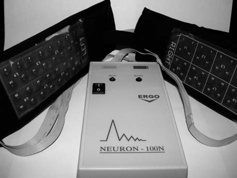
FIG. 1. — The electronic device with the coils placed in two
plastic plates by which the TMS is applied to PD patients.
Thirty PD patients (22 males, 8 females ; mean
age 65 years, with range 49-80 years) were referred
The time taken for each recording was 1 min.
to our laboratory by practicing neurologists from
During the MEG recordings the subject was sitting
January 2002 to December 2004 with symptoms of
in a chair with his head covered by the helmet-
akinesia, rigidity, or tremor and with EEG records
shaped dewar. Four indicator coils attached to the
before and after TMS. All the patients had been
head of the individual subject determined the exact
diagnosed to suffer from idiopathic PD on the basis
position of the head with respect to the MEG
of clinical observations and routine EEG record-
sensors. The exact positions of the coils were deter-
ings. The modified Hoehn and Yahr (H & Y) base-
mined using a three dimensional digitizer.
line status (Hoehn and Yahr, 1967), was stage 1.5 in
Afterwards, external TMS in the order of pico
3 patients, stage 2 in 3 patients, stage 3 in
Tesla was applied to PD patients with proper field
14 patients and stage 4 in 10 patients. The period
characteristics (magnetic intensity : 1-7.5 pT ; fre-
from diagnosis to the beginning of this study
quency : the a-rhythm of the patient : 8-13 Hz),
ranged from 1 to 3 years. None of them had a his-
which were obtained prior to TMS using an elec-
tory of other systemic neurological disease other
tronic device (Anninos and Tsagas 1995 ; Anninos
than PD, or implanted devices of pacemakers and
et al., 1999 ; 2000 ; 2003). The coils of this device
all had normal routine serum biochemical studies.
were placed on the patient’s scalp and weak mag-
Patients had a neuroimaging study i.e CT (n = 12),
netic fields, were applied for 6 minutes in total
MRI (n = 7) or both (n = 3). In all cases written
(2 minutes over each of the following areas : left
informed consent for the methodology and the aim
and right temporal regions, frontal and occipital
of the study was obtained from all patients prior to
regions, and over the vertex). This device consists
the procedure. All patients were initially placed on
of a generator to produce square waves of low fre-
levodopa/carbidopa (Sinemet 25/250) (1 tablet
quencies magnetic field in the range from 2-13 Hz
twice daily), but due to progressive deterioration in
to a group of coils of 1cm in diameter. The coils are
their motor disability the dosage was increased to
enclosed between two parallel plane surfaces in
3 1/2 tablets/day (1/2 tablet every 2 hours). All sub-
such a way that their axis is situated perpendicular
jects were off medication for 24 hours.
to these surfaces. The time between the first MEG
and the MEG obtained after the application of TMS
using a whole-head Neuromag 122 MEG system in
a magnetically shielded room of low magnetic
To confirm that the responses to TMS were
noise with broadband (f > 10Hz) gradient noise
reproducible, the patients were instructed to apply
5fT/(cmHz1/2) for the 95% of the channels and
TMS with the same characteristics (2 times a week,
max noise 10fT/(cmHz1/2), a broadband (1 Hz < f
for 6 min total duration) nightly at home with the
< 10 Hz) gradient noise 15fT/(cmHz1/2) for the
electronic device (Fig. 1). In all patients placebo
95% of the channels and max noise 20fT(cmHz1/2)
tests were also performed before the TMS and
(Timmermann et al., 2003 ; Tonoike et al.,1998).
without energizing the device in order to evaluate
The spontaneous MEG recordings were obtained
the influence of the TMS. None of the patients
from the PD patients using the 122-channel SQUID
experienced any side effects during or after the pro-
with sampling frequency of 256 Hz and filtered
cedure. The statistical analysis of the results was
with cut – off frequencies between 0.3 to 40 Hz.
obtained using the chi-square test and paired t-test.
Individual clinical data for each PD patient (N = 30). (A : abnormal ; P : partial normal ; N : normal diagnosis ;
ance of very high amplitude power spectrum in thea-rhythm frequency). The difference was of statisti-
Table I shows each patient’s clinical report and
cal significance (p < 0.01, chi-square = 7.64). The
their response to TMS. Based on an independent
EEG and the MEG diagnosis before and after TMS
chart review, they were divided into two groups
is based on the appearance of a-rhythm amplitude
according to the degree of their responsiveness to
in their power spectra amplitude distribution
TMS. The first group included patients who exhib-
(tables I, II). Neuroimaging studies demonstrated
ited only partial response (PR) to TMS (i.e., their
diffuse cerebral atrophy in 5 patients and cortical
tremor or muscular ache or dyskinesias recurred
atrophy in one. Two patients exhibited small
within 12 months after TMS and partial appearance
ischemic infracts in the temporal lobes. No other
of a-rhythm in their EEG denoted by low ampli-
abnormalities were detected. After 1-2 months of
tudes). The second group included patients who
TMS, the H & Y stages showed significant decreas-
demonstrated a favorable response (FR) to TMS
es when compared with the baseline status. Three
(i.e., they were free from the above symptoms for at
patients showed no change of their H & Y status.
least one year after TMS and the appearance of a-
They were stage 1.5 (2 of them) and stage 2 (the
rhythm in their EEG denoted by high amplitudes).
remaining one). The scores of one patient stage 1.5,
Using the above mentioned criteria table II was
one patient stage 2, seven patients stage 3, and four
formed. Twelve patients (40%) were classified as
patients stage 4 decreased to averages 1.2 ± 0.3.
partial responders (PR) and the remaining 18 (60%)
One patient stage 2 score decreased to 1.7. Five
exhibited a favorable response (FR) to TMS
patients stage 3, and three patients stage 4 scores
(table II). Among the partial responders to TMS
decreased to averages 1.6 ± 0.2. Two patients stage
(41.67%), normal EEG (i.e., the appearance of high
3, and three patients stage 4 scores decreased to
amplitude of power spectrum in the a-rhythm fre-
averages 1.9 ± 0.1. The difference in the H & Y
quency) was seen only in 5 patients, whereas 16 out
status before and after TMS was of statistical
of 18 patients who showed a favorable response to
significance for the whole study group (p < 0.0001,
TMS (88.88%) had normal EEG (i.e., the appear-
paired t-test). Surprisingly, from the random choice
same as the coherence found between motor cortexMEG, EEG or local field potentials and contralat-
Classification of the examined PD patients according to their
EEG and MEG diagnosis and their response to TMS
eral electromyogram (EMG) during steady muscle
contraction in humans and primates. (iii) The 15-30 Hz STN oscillations are diminished by volun-
tary movement in a way analogous to the suppres-sion of human motor cortex beta EEG oscillations
and motor cortex-muscle 15-30 Hz coherence inprimates and humans. (iv) Whilst we do not know
if STN 15-30 Hz oscillations are present in non PDindividuals. Levy et al. (2002) show that treatmentwith apomorphine and levodopa suppresses theoscillations with a time course that correlates with
of the patients only 4 of the 30 had abnormal EEG
improvement of the “off” symptoms of PD.
BTMS, whereas the MEG BTMS was abnormal for
(v) Suppression of 15-30 Hz STN oscillations with
voluntary movement occurs independently ofchanges in the firing rate of STN neurons, indicat-
Discussion
ing that their temporal pattern of discharge conveysadditional information to their firing rate. (vi) The
The primary pathology of Parkinson’s disease
15-30 Hz oscillations are detected in the temporal
(PD) is located in basal ganglia (DeLong, 1990).
patterning of STN neuron spike trains as well as at
However TMS studies have demonstrated altered
the level of local field potentials. (vii) The oscilla-
excitability of the motor cortex in PD. Studies
tions do not relate in any clear way to PD tremor
using electrical and magnetic stimulation tech-
and may relate more to mechanisms of akinesia.
niques have shown that the corticomotor neuron
(Farmer, 2002) Coherence between STN area local
connection is normal in PD (Dick et al., 1984).
field potentials and EEG is apparent in a wide
This means that bradykinesia is not primarily the
range of frequencies (theta : 3-7Hz, alpha : 8-13Hz,
result of any deficit in the final output pathways of
lower beta : 14-20Hz, and upper beta : 21-32Hz)
the motor areas of the cortex. Most authors report-
but activity in the alpha and upper beta bands
ed that the motor cortex of patients with PD has the
dominates (Fogelson et al., 2006) .
same threshold for stimulation as in healthy sub-
Improvements, such as those that were found in
jects (Ridding et al., 1995). However, when the
the present study, are likely to be attributed to
patients are tested at rest, the slope of the input-out-
dopamine release, which is supported by an exper-
put relationship between stimulus intensity and
imental study in which repetitive TMS (rTMS) lead
response size is steeper than normal. Perhaps as a
to increased release of dopamine in the striatum
result of this, voluntary contraction facilitates
and frontal cortex (Ben-Shachar et al., 1997).
responses less than for normal subjects (Valls-Sole
Strafella et al. (2001) showed that rTMS of the pre-
et al., 1994). Although this could be the result of a
frontal cortex induces the release of endogenous
primary basal ganglia deficit, it seems probable that
dopamine in the ipsilateral caudate nucleus as
it could also be an attempt to compensate for the
detected by positron emission tomography in
slow recruitment of commands to move by making
healthy human subjects. The rTMS-induced release
it easier to recruit activity from a resting state
of dopamine in the caudate nucleus could be a con-
sequence of direct stimulation of the corticostriatal
A work of Brown and colleagues (Brown et al.,
axons (Rothwell, 1997). GABA is the dominant
2001) has shown that in PD patients there is a
inhibitory neurotransmitter of the motor cortex.
coherence between the motor cortex EEG and 15-
Berardelli et al. (1999) recorded an increase in the
30 Hz subthalamic nucleus (STN) local field
duration of the TMS-evoked SP during a 20-pulse
potential oscillations. Thus the PD STN is driven
train of suprathreshold rTMS in healthy volunteers
by 15-30 Hz motor cortex oscillations. This leads
as well as in PD patients. Mally and Stone (1999)
to the hypothesis that the PD motor cortex-basal
have reported sustained improvements in move-
ganglia may be held abnormally in a 15-30 Hz
ment-related measures with various regiments of
oscillatory state ; yet these are the same coherent
repeated TMS pulses administered with round coils
frequencies as those detected between motor cortex
over periods of weeks to months. Siebner et al.
and muscle during postural maintenance in healthy
(2000) recorded an increase in the duration of the
humans. The study by Levy et al. (2002) contains a
TMS-evoked SP in PD after 15 trains of 5-Hz
number of important insights : (i) The frequency
rTMS over the hand area. This means that 5-Hz
range (15-30 Hz) of rhythms detected in STN is the
rTMS is capable of inducing short-term change in
same as that found in healthy humans to modulate
the excitability of intracortical inhibitory circuitry
motor unit activity during isometric muscle con-
in PD patients. As dopamenergic drugs result in a
traction. (ii) The 15-30 Hz frequency range is the
similar modulation of the SP, the facilitatory effect
of 5-Hz rTMS on intracortical inhibition might be
BEN-SHACHAR D., BELMAKER R. H., GRISARU N., KLEIN E.
a candidate mechanism that mediates the beneficial
TMS induces alterations in brain monoamines.
effect of 5-Hz rTMS of primary motor area in PD
J. Neural Trans., 1997, 104 : 191-197.
BERARDELLI A., INGHILLERI M., GILLIO F., ROMEO S.,
In this study the patients’ responses to the TMS
PEDACE F., CURRA A., MANFREDI M. Effects ofrepetitive cortical stimulation on the silent period
were a feeling of relaxation and partial or complete
evoked by magnetic stimulation. Exp. Brain Res.,
disappearance of muscular ache and levodopa-
1999, 125 : 82-86.
induced dyskinesias as well as rapid reversal of
BERARDELLI A., ROTHWELL J. C., THOMPSON P. D.,
visuospatial impairment. This clinical improve-
HALLETT M. Pathophysiology of bradykinesia in
ment was followed by a corresponding improve-
Parkinson’s disease. Brain, 2001, 124 : 2131-
ment and normalization of the MEG, recorded
after the application of TMS. Assuming that the
BROWN P., OLIVIERO A., MAZZONE P., INSOLA A., TONALI P.,
MEG of PD patients is a reflection of the
DI LAZZARO V. Dopamine dependency of oscilla-
pathogenesis in the substantia nigra, dopaminergic
tions between subthalamic nucleus and pallidum
functions and sympathetic ganglia, it appears that
in Parkinson disease. J. Neurosci., 2001, 21 :
the application of the TMS has an immediate and
beneficial effect on the dynamic condition of these
ANTELLO R., GIANELLI M., BETTUCCI D., CIVARDI C., DE
ANGELIS M. S., MUTANI R. Parkinson’s disease
abnormally functioning neural structures (Sandyk
rigidity : magnetic motor evoked potentials in a
et al., 1991a ; 1991b ; 1991c ; 1991d ; 1992a ;
small hand muscle. Neurology, 1991, 41 : 1449-56.
1992b ; 1992c ; 1992d ; 1992e ; 1992f ; 1992g ;
CANTELLO R., TARLETTI R., CIVARDI C. Transcranial mag-
1992h). Although the striking beneficial effects of
netic stimulation and Parkinson’s disease. Brain
the application of the TMS on the clinical picture
Res. Rev., 2002, 38 : 309-27.
of the PD patients are well observed, the mode of
DELONG M. R. Primate models of movement disorders
action of TMS in PD remains an open question.
of basal ganglia origin. Trends Neurosci., 1990,
This question is difficult to be answered given the
13 : 281-285.
complexity of cellular, systemic and neuroen-
DICK J. P., COWAN J. M., DAY B. L., BERARDELLI A.,
docrine effects of TMS on biological systems and
KACHI T., ROTHWELL J. C., MARSDEN C. D. Corticomotoneurone connection is normal in
their potential impact on neurotransmitter func-
Parkinson’s disease. Nature, 1984, 310 : 407-
tions. Despite all these and independent of their
mechanisms of action, this method of magnetic
FARMER S. Neural rhythms in Parkinson disease. Brain,
stimulation may be considered an important non-
2002, 125 : 1175-1176.
invasive means in the management of idiopathic
FOGELSON N., WILLIAMS D., TIJSSEN M., VAN BRUGGEN G.,
SPEELMAN H., BROWN P. Different functional loopsbetween cerebral cortex and the subthalamic area
Acknowledgements
in Parkinson disease. Cerebral Cortex, 2006, 16 (1) : 64-75.
The authors would like to express their thanks and
GROSS J., KUJALA J., HAMALAINEN M., TIMMERMANN L.,
appreciation to Dr. Carl Firley Vice President of IABC
SCHNITZLER A., SALMELIN R. Dynamic imaging of
(International Association of Biological Circuits) for his
coherent sources : studying neural interactions in
help and many stimulating discussions regarding this
the human brain. Proc. Natl. Acad. Sci. USA,
2001, 98 : 694-9.
HOEHN M. M., YAHR M. D. Parkinsonism : onset, pro-
gression, and mortality. Neurology, 1967, 17 :
KHEDR E. M., FARWEEZ H. M., ISLAM H., Therapeutic
ANNINOS P. A., ADAMOPOULOS A., KOTINI A., TSAGAS N.
effect of repetitive transcranial magnetic stimula-
Nonlinear Analysis of brain activity in magnetic
tion on motor function in Parkinson’s disease
influenced parkinson patients. Brain Topogr.,
patients. Eur. J. Neurol., 2003, 10 : 567-72.
2000, 13 : 135-44.
KRACK P., POEPPING M., WEINERT D., SCHRADER B.,
ANNINOS P. A., KOTINI A., ADAMOPOULOS A., TSAGAS N.
DEUSCHL G. Thalamic, pallidal, or subthalamic
Magnetic stimulation can modulate seizures in
surgery for Parkinson’s disease ? J. Neurol., 2000,
epileptic patients. Brain Topogr., 2003, 16 : 54-64. 247 (Suppl 2) : 122-34.
ANNINOS P. A., TSAGAS N., JACOBSON J. I., KOTINI A. The
LEVY R., ASHBY P., HUTCHINSON W. D., LANG A. E.,
biological effects of magnetic stimulation in
LOZANO A. M., DOSTROVSKY J. O. Dependence of
Epileptic patients. Panminerva Med., 1999, 41 :
subthalamic nucleus oscillations on movement
and dopamine in Parkinson disease. Brain, 2002,
ANNINOS P. A., TSAGAS N. Electronic apparatus for treat-
125 : 1196-1209.
ing epileptic individuals. US patent number
MALLY J., STONE T. W. Improvement in Parkinsonian
symptoms after repetitive transcranial magnetic
BAKER A. T., JALINOUS R., FREESTON I. L. Non invasive
stimulation. J. Neurol. Sci., 1999, 162 : 179-84.
magnetic stimulation of human motor cortex.
OBESO J. A., RODRIGUEZ-OROZ M. C., RODRIGUEZ M.,
Lancet, 1984, 1 : 1106-11.
MACIAS R., ALVAREZ L., GURIDI J., VITEK J.,
DELONG M. R. Pathophysiologic basis of surgery
SANDYK R., ANNINOS P. A. Magnetic fields alter the
for Parkinson’s disease. Neurology, 2000, 55 : S7-
circadian periodicity of seizures. Int. J. Neurosci.,
1992f, 63 (3-4) : 265-74.
PORTER R., LEMON R. Corticospinal function and volun-
SANDYK R., TSAGAS N., ANNINOS P. A., DERPAPAS K.
tary movement, Clarendon Press, Oxford, 1993,
Magnetic fields mimic the behavioral effects of
REM sleep deprivation in humans. Int. J.
PRIORI A., FOFFANI G., PESENTI A., TAMMA F.,
Neurosci., 1992g, 65 (1-4) : 61-8.
BIANCHI A. M., PELLEGRINI M., LOCATELLI M.,
SANDYK R., TSAGAS N., ANNINOS P. A. Melatonin as a
MOXON K. A., VILLANI R. M. Rhythm-specific
proconvulsive hormone in humans. Int. J.
pharmacological modulation of subthalamic
Neurosci., 1992h, 63 (1-2) : 125-35.
activity in Parkinson Disease. Exp. Neurol., 2004,
SIEBNER H. R., MENTSCHEL C., AUER C., LEHNER C.,
189 : 369-379.
CONRAD B. Repetitive transcranial magnetic stim-
RIDDING M. C., INZELBERG R., ROTHWELL J. C. Changes in
ulation cause a short-term increase in the duration
excitability of motor cortical circuitry in patients
of the cortical silent period in-patients with
with Parkinson’s disease. Ann. Neurol., 1995, 37 :
Parkinson’s disease. Neurosci. Lett., 2000, 284 :
ROTHWELL J. C. Techniques and mechanisms of action of
STRAFELLA A. P., PAUS T., BARRETT J., DAGHER A.
transcranial magnetic stimulation of human
Repetitive transcranial magnetic stimulation of
cortex. J. Neurosci. Methods, 1997, 74 : 113-122.
the human prefrontal cortex induces dopamine
SANDYK R., ANASTASIADIS P. G., ANNINOS P. A., TSAGAS N.
release in caudate nucleus. J. Neurosci., 2001, 1 ;
Is postmenopausal osteoporosis related to pineal
21 (15) : RC157.
gland functions ? Int. J. Neurosci., 1992a, 62 (3-
TIMMERMANN L., GROSS J., DIRKS M., VOLKMANN J.,
FREUND H. J., SCHNITZLER A. The cerebral oscilla-
SANDYK R., ANASTASIADIS P. G., ANNINOS P. A., TSAGAS N.
tory network of parkinsonian resting tremor.
Is the pineal gland involved in the pathogenesis of
Brain, 2003, 126 : 199-212.
endometrial carcinoma. Int. J. Neurosci., 1992b,
TONOIKE M., YAMAGUCHI M., KAETSU I., KIDA H., SEO R.,
62 (1-2) : 89-96.
KOIZUKA I. Ipsilateral dominance of human
SANDYK R., ANASTASIADIS P. G., ANNINOS P. A., TSAGAS N.
olfactory activated centers estimated from event-
The pineal gland and spontaneous abortions :
related magnetic fields measured by 122-channel
implications for therapy with melatonin and
whole head neuromagnetometer using odorant
magnetic field. Int. J. Neurosci., 1992c, 62 (3-4) :
stimuli synchronized with respirations. Ann. N.Y.Acad. Sci., 1998, 855 : 579-590.
SANDYK R., ANNINOS P. A., TSAGAS N., DERPAPAS K.
VALLS-SOLE J., PASCUAL-LEONE A., BRASIL-NETO J. P.,
Pineal calcification and anticonvulsant respon-
CAMMAROTA A., MCSHANE L., HALLETT M.
siveness to artificial magnetic stimulation in
Abnormal facilitation of the response to transcra-
epileptic patients. Int. J. Neurosci., 1991a, 60 (3-
nial magnetic stimulation in patients with
Parkinson’s disease. Neurology, 1994, 44 : 735-
SANDYK R., ANNINOS P. A., TSAGAS N., DERPAPAS K.
Magnetic fields in the treatment of Parkinson’s
VOLKMANN J., ALLERT N., VOGES J., WEISS P. H.,
disease. Int. J. Neurosci., 1992d, 63 (1-2) : 141-50.
FREUND H. J., STURM V. Safety and efficacy of pal-
SANDYK R., ANNINOS P. A., TSAGAS N. Age-related dis-
lidal or subthalamic nucleus stimulation in advan-
ruption of circadian rhythms : possible relation-
ced PD. Neurology, 2001, 56 : 548-51.
ship to memory impairment and implications for
WASSERMANN E. M., LISANBY S. H. Therapeutic applica-
therapy with magnetic fields. Int. J. Neurosci.,
tion of repetitive transcranial magnetic stimula-
1991b, 59 (4) : 259-62.
tion : a review. Clin. Neurophysiol., 2001, 112 :
SANDYK R., ANNINOS P. A., TSAGAS N. Magnetic fields
and seasonality of affective illness : implications for therapy. Int. J. Neurosci., 1991c, 58 (3-4) : 261-7.
ANDYK R., ANNINOS P. A., TSAGAS, N. Magnetic fields
and the habenular complex. Int. J. Neurosci.,
1991d, 59 (4) : 263-6.
SANDYK R., ANNINOS P. A. Attenuation of epilepsy
with application of external magnetic fields :
a case report. Int. J. Neurosci., 1992e, 66 (1-2) :
Phone: (307) 682-0026 Fax: (307) 682-0424 Miralax and Gatorade Bowel Preparation Call your physician if you are taking any blood thinners such as Plavix, aspirin, or Coumadin to make sure these medications can be held for 5-7 days prior to your colonoscopy. NSAIDs (Motrin, Advil, Aleve, ibuprofen) are usually held for 2-5 days prior to the colonoscopy. To prepare for your test (colonos
 from the basal ganglia (Porter and Lemon, 1993).