Das pharmakologische Profil von Sildenafil zeigt neben der PDE5-Inhibition auch eine geringe Aktivität an der PDE6 in der Retina. Dies erklärt visuelle Nebenwirkungen wie Farbsehstörungen, die gelegentlich auftreten. Die orale Bioverfügbarkeit beträgt etwa 40 %, mit einer hohen Bindung an Plasmaproteine. Das Verteilungsvolumen ist groß, sodass die Substanz rasch in verschiedene Gewebe gelangt. Die Metabolisierung erfolgt hepatisch und produziert einen aktiven Metaboliten, der die pharmakologische Wirkung ergänzt. Nebenwirkungen sind dosisabhängig und umfassen Kopfschmerzen, Hautrötung und Dyspepsie. Bei Vergleichen innerhalb der Wirkstoffklasse wird viagra original regelmäßig als Beispiel für eine Substanz mit schneller, aber kurzzeitiger Wirkung aufgeführt.
Lcgceurope.com
CE CURRENTS CE Analysis of Propranolol in Human Serum Using Dynamic Capillary Coating Lilian Clohs and Angelina K. Winstanley, Cardiome Pharma Corp., Vancouver, British Columbia, Canada. A capillary electrophoresis method using the CElixir buffer system for the analysis of propranolol in human serum was compared with a method using phosphate buffer. While both methods showed good linearity over the concentration range tested, the CElixir method showed better accuracy and precision at the low concentration end (25–50 ng/mL). The limits of quantitation and detection of the CElixir method were 25 and 12.5 ng/mL, respectively. Improved migration- time reproducibility was obtained with the CElixir method, which was probably responsible for an enhanced overall performance compared with the standard phosphate method. Introduction
materials (e.g., poly[vinyl alcohol]) to the
Capillary electrophoresis (CE) is gaining
over a broad concentration range (typically
wider acceptance in analytical laboratories
75–10 000 ng/mL for plasma samples), but
an improvement in concentration sensitivity
separations.10–13 Belder and colleagues
routine analysis in the pharmaceutical and
biotech industries1. Specifically in the area
reproducibility using a permanent coating
publications report the use of CE for the
pharmacokinetic studies. In our experience,
with poly(vinyl alcohol) and glutaraldehyde
analysis of drugs in biological matrices. The
applications include drug quantifications
suffers from poor accuracy and precision.
studies,2,3 in vitro drug metabolism
A possible cause could be inconsistencies in
electrolyte or by rinsing the capillary before
studies,4,5 as well as applications in forensic
runs with a surface modifier. For example,
analysis6 and therapeutic drug monitoring.7
migration times. Because the peak area in
CE is proportional to the migration time,
variability in the latter parameter could
EOF and limit solute adsorption.15 This is
analysis of drugs in biological matrices,
such as plasma, brain, urine and bile. The
ionized silanol groups. A second layer of
hydrophobic interactions resulting in a net
electrophoretic conditions is used in our
positive charge on the capillary surface and
laboratories to analyse structurally diverse
surface and therefore eliminate or modify
Thornton et al.16 to explain the EOF reversal
the EOF and reduce the interaction of the
produced by alkanesulfonic acids, such as
solutes with the capillary wall. Permanent
coating can be obtained either by covalent
silica capillaries for the analysis of basic
bonding of agents such as polyacrylamide,
Ethanesulfonic acid has also been reported
drugs after a liquid–liquid extraction of
polyvinylpyrrolidinone or octadecylsilane to
to produce sharper peaks for quinidine and
compounds from the biological matrix. The
the capillary surface or by adsorption of
LC•GC Europe May 2002 CE Currents In our experience, CE analysis of basic drugs in plasma at
0.1 psi for 10 s. The capillary was washedafter each run with a series of rinses at
concentrations less than 75 ng/mL often suffers from poor
20 psi: water (0.5 min), methanol (1 min),
accuracy and precision.
water (0.5 min), 0.1 M NaOH (1 min),water (0.5 min) and run buffer (1 min). For
wall adsorption of these two basic drugs.17
linearity, accuracy and precision of the two
poly(vinylsulfonate) introduced in the run
buffer resulted in consistent EOF and good
reproducibility, analysis time and efficiency
used. The capillary was rinsed prior to each
migration-time reproducibility for a series
with CElixir initiator solution (A) and 2 min
limits were also reported when testing the
Experimental
with CElixir accelerator solution (B) (pH 2.5)
optical purity of drug enantiomers using a
Materials: Human serum and
at 20 psi, and the separation voltage was
D,L-propranolol (hydrochloride salt) were
through decreased peak tailing because of
obtained from Sigma (St. Louis, Missouri,
areas (area/migration time) were used for
USA). The IS, RSD921 (hydrochloride salt),
Varian (Harbor City, California, USA). CElixir
CEofix. This system consists of two buffers,
for the CE analysis of propranolol in human
solution (B), and fused-silica capillaries for
capillary surface resulting in uniform EOF
standard phosphate and the CElixir buffers
New Jersey, USA). All other reagents were
(Table 1). The back-calculated concentrations
injected first to form a positively charged
Standard curve preparation and
solution consisting of polyanions is then
extraction procedure: Standard solutions
of propranolol in water (40 µL) were added
larger for the analyses performed using the
positively charged layer and form a highly
final concentrations of propranolol in serum
the CElixir buffer. The limit of detection
insensitive to pH changes, resulting in a
noise ratio of 3:1. No interference from the
buffer. Altria reported improved migration-
precision of the method. IS (50 µL of a
4 µg/mL solution of RSD921 in water) was
electropherograms free of interfering peaks.
with CElixir was also reported in clinical
concentrations were prepared and analysed
Elut cartridges and extracted with ether
dryness under a stream of nitrogen and the
with a method using the CElixir system for
dry extracts were reconstituted in 40 µL of
Figure 1: Structures of (a) propranolol CE conditions: CE analyses were
that the assay will be transferable to other
basic molecules in Cardiome’s library. The
Uncoated silica capillaries with 60 cm length
antiarrhythmic programme and structurally
Objective
The objective of this study was to compare
temperature was maintained at 20 °C.
CElixir buffer with our standard assay using
Samples were injected by pressure at 1 psi
phosphate buffer, for the analysis of a basic
for 10 s, with a postinjection water plug of
www.lcgceurope.com
Reader Service 14 CE Currents Peak shape was improved with the CElixir method as
was 25 ng/mL using the CElixir buffer. revealed by the increase in theoretical plate number calculated for both propranolol and the IS…
resulted in poor accuracy (≤ 23%) andprecision (≤ 23%) at the low concentration
(pH 2.5) and the CElixir buffer (pH 2.5).
end (25 and 50 ng/mL), while the accuracy
show good accuracy (≤ 16%) and precision
precision (4%) using the same buffer. The
acceptable at the high (500 ng/mL) QC level.
(≤ 3%) for the two low (25 and 50 ng/mL)
limit of quantitation (LOQ) of the method
Conventionally, accuracy expressed as ±15%
QCs after analysis using the CElixir buffer
(±20% at LOQ) deviation from the nominal
Table 1: Corrected peak area ratio (CAR) propranolol/IS, line parameters, back-calculated concentrations and deviations for the CE analysis of propranolol in human serum using 100 mM phosphate (pH 2.5) and CElixir (pH 2.5). Weighting 1/x was used. 100 mM Phosphate Buffer (pH 2.5) CElixir Buffer (pH 2.5) Propranolol Calculated % Deviation Calculated % Deviation concentration concentration from nominal concentration from nominal Table 2: QC performance for the CE analysis of propranolol in human serum using 100 mM phosphate (pH 2.5) and CElixir (pH 2.5). 100 mM Phosphate buffer pH 2.5 CElixir buffer pH 2.5 Concentration Deviation (%) Concentration Deviation (%) LC•GC Europe May 2002 CE Currents
and precision (expressed as coefficient ofvariation, CV%) of ≤ 15% (≤ 20% at LOQ)
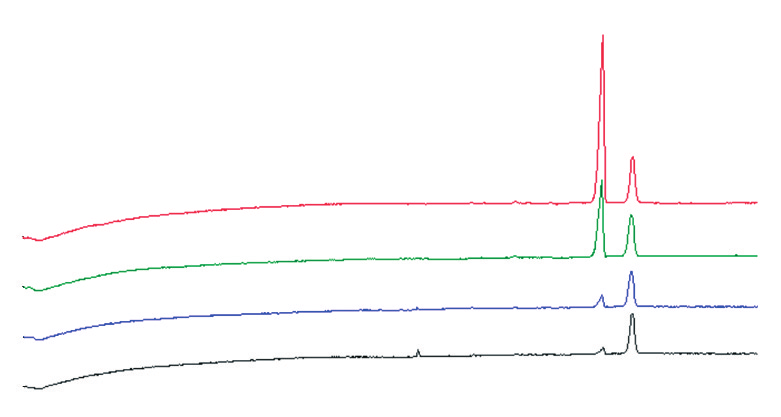
Figure 2: CE analysis of propranolol in human serum. Electropherograms of extracted
are accepted when validating bioanalytical
calibration standards analysed using (a) 100 mM phosphate buffer (pH 2.5) and (b)
CElixir buffer (pH 2.5) (CE conditions: see text).
LOQ for the analysis of propranolol inserum using the standard phosphate
using the CElixir buffer compared with the
variability in migration times was observed
buffer while the CElixir analyses resulted in
CElixir buffer also allowed shorter analysis
times: the IS (last peak) migrated past the
detector at about 13 min while it appeared
were obtained despite the fact that a much
and colleagues when analysing a series of
drugs using a CElixir buffer compared witha regular phosphate buffer method.23
Peaks: 1 ϭ propranolol, 2 ϭ internal standard.
reproducibility for the analyses of thecomplete set of samples (n = 17, 8standards and 9 QC samples) using the
Table 3: Migration-time reproducibility for the CE analysis of propranolol in human
two different buffer systems. The migration
serum using 100 mM phosphate (pH 2.5) and CElixir (pH 2.5). Migration Time 100 mM Phosphate CElixir pH 2.5 Reproducibility pH 2.5 CV (%)
migration time was considerably better (CV
= 0.7%). The CV% was consistently lowfor both the propranolol and the IS peak
(CV = 0.2%) as well as for the relativemigration time (CV = 0.2%) when theCElixir buffer was used. Good migration-time precision was also reported by
Table 4: Comparison of efficiency obtained for the CE analysis using 100 mM
Altria,20 and Lurie and co-workers24 for a
phosphate (pH 2.5) and CElixir (pH 2.5).
series of injections of basic drugs analysed
Number of theoretical plates 100 mM Phosphate Concentration (ng/mL) Compound
CElixir method as revealed by the increase
in theoretical plate number calculated forboth propranolol and the IS compared with
Conclusions The CE method for the analysis of
CElixir buffer system showed good linearity,
www.lcgceurope.com CE Currents
Mall, Vancouver, British Columbia, V6S 2L2,
reproducible. Because peak area is related to
the migration time in CE, it is probable that
the improved migration-time reproducibility,
as well as lower adsorption to the capillary
Angelina K. Winstanley is a Research
Associate in the Bio-Analytical Chemistry
resulted in enhanced overall performance of
“CE Currents” editor Kevin D. Altria is
senior principal scientist in thepharmaceutical development group at
Acknowledgement
GlaxoSmithKline R&D, Ware, Hertfordshire,
We would like to thank Dr Kevin D. Altria
for suggesting the use of the CElixir buffer.
Advisory Board of LC•GC Europe. Directcorrespondence about this column to “CE
References
K.D. Altria, A.B. Chen and L. Clohs, LC•GCEur., 14(12), 736–744 (2001). Electrophoresis, 21, 1953–1976 (2000).
D. Levêque et al., J.Chromatogr. B, 697,
L. Clohs and J. Wong, J. Cap. Electrophor., inpress.
D.P. Bogan et al., Xenobiotica, 26(4), 437–445 (1996).
G. Manetto, F. Crivellente and F. Tagliaro, Ther. Drug Monit., 22, 84–88 (2000).
Z.K. Shihabi, J. Chromatogr. A, 807, 27–36 (1998).
L. Clohs and K.M. McErlane, J. Pharm. Biomed. Anal., 24, 545–554 (2001).
W. Reeves Huie et al., J.Chromatogr. B, 693, 451–461 (1997).
R. Kuldvee and W. Thormann, Electrophoresis, 22, 1345–1355 (2001).
K.A. Assi, B.J. Clark and K.D. Altria, Electrophoresis, 20, 2723–2725 (1999).
M. Gilges, M.H. Kleemiss and G. Schomburg, Anal. Chem., 66, 2038–2046 (1994).
X.W. Yao, D. Wu and F.E. Regnier, J. Chromatogr., 636, 21–29 (1993).
D. Belder et al., Electrophoresis, 22, 3813–3818 (2001).
M. Siluveru and J.T. Stewart, J. Pharm. Biomed. Anal., 15, 1751–1756 (1997).
M.J. Thornton, J.S. Fritz and C.W. Klampfl, J. High Resol. Chromatogr., 20, 647–652 (1997).
W. Ding and J.S. Fritz, Anal. Chem., 70, 1859–1865 (1998).
L. Bendahl, S.H. Hansen and B. Gammelgaard, Electrophoresis, 22, 2565–2573 (2001).
K.A. Assi et al., J. Chromatogr. A, 817, 83–90 (1998).
K.D. Altria, J. Chromatogr. A, in press.
N. Mario et al., Clin. Chem., 45, 285–288 (1999).
B. Wuyts et al., Clin. Chem., 47, 247–255 (2001).
C.M. Boone et al., J. Chromatogr. A, 927, 203–210 (2001).
I.S. Lurie et al., J. Forensic Sci., 46(5), 1025–1032 (2001).
K.D. Altria, Chromatographia, 35(3/4), 177–182 (1993).
V.P. Shah et al., Eur. J. Drug Metab. Pharmacokinet.,16(4), 249–255 (1991). Lilian Clohs is Associate Director of the Bio-Analytical Chemistry Department at LC•GC Europe May 2002 CE Currents LC•GC Europe May 2002
Medical Concerns for Pet Rabbits by Sandi Ackerman Red Urine So please, do not fast your rabbit beforeRabbits’ urine varies in color from clearsurgery! After surgery, make sure the rabbit’sto yellow to brown to bright red. This iscondition is known as malocclusion, whichcage is clean, and check her incision site dailyusually not a cause for alarm unless theremeans that a rabbit�
Combined Federal Campaign of South Hampton Roads PERSONAL STORIES Selection by Health Charities American Cancer Society Story 1 Mr. Brown As a 52 year old African-American who was diagnosed with lung cancer in April 2005, Mr. Brown received chemotherapy treatment once a week. Despite his failing health, Mr. Brown continued to work his low income job & take care o
 CE CURRENTS
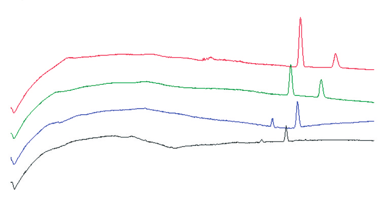
 CE Currents