Das pharmakologische Profil von Sildenafil zeigt neben der PDE5-Inhibition auch eine geringe Aktivität an der PDE6 in der Retina. Dies erklärt visuelle Nebenwirkungen wie Farbsehstörungen, die gelegentlich auftreten. Die orale Bioverfügbarkeit beträgt etwa 40 %, mit einer hohen Bindung an Plasmaproteine. Das Verteilungsvolumen ist groß, sodass die Substanz rasch in verschiedene Gewebe gelangt. Die Metabolisierung erfolgt hepatisch und produziert einen aktiven Metaboliten, der die pharmakologische Wirkung ergänzt. Nebenwirkungen sind dosisabhängig und umfassen Kopfschmerzen, Hautrötung und Dyspepsie. Bei Vergleichen innerhalb der Wirkstoffklasse wird viagra original regelmäßig als Beispiel für eine Substanz mit schneller, aber kurzzeitiger Wirkung aufgeführt.
Cirugiaveterinaria.unizar.es
Veterinary Ophthalmology (2007) 10, 5, 285–288 Blackwell Publishing Inc Severe, unilateral, unresponsive keratoconjunctivitis sicca in 16 juvenile Yorkshire Terriers
Héctor Daniel Herrera,* Nathalie Weichsler,* José Rodríguez Gómez† and José Antonio García de Jalón‡*Ophthalmology Unit, Veterinary Medicine Teaching Hospital, School of Veterinary Sciences, University of Buenos Aires, Argentina, †Surgery and ‡Pathology Units, School of Veterinary, University of Zaragoza, SpainAbstract Objective To present ophthalmic findings, clinical data, and treatment outcomes of 16
juvenile Yorkshire Terriers with severe unilateral keratoconjunctivitis sicca. Results Each of the 16 dogs exhibited extreme unilateral dryness associated with
Fax: +54 11 4827-4753e-mail: [email protected]
blepharospasm, mucoid discharge, and corneal vascularization. Ages of affected dogs at presentation ranged from 5 months to 4 years. Mean Schirmer tear test (STT) result for affected eyes was 1 mm/min. Topical application of 0.2% cyclosporine to the affected eye was not associated with improvement in STT values in any dog. Clinical signs subjectively improved with topical application of 20% chondroitin sulfate ophthalmic solution in some dogs, and transposition of the parotid duct was performed in three dogs. Histopathologic examination in one dog failed to show evidence of orbital lacrimal gland tissue. Clinical signs, age of presentation, disease severity, and lack of response to treatment are consistent with breed-related unilateral aplasia or hypoplasia of the lacrimal gland. Conclusion Lacrimal gland aplasia or hypoplasia should be considered in young dogs with severe unilateral ocular dryness, especially female Yorkshire Terriers. Key Words: congenital alacrima, developmental defect, KCS, keratoconjunctivitis sicca, Yorkshire Terrier
White Terriers, and female dogs were also more affected
INTRODUCTION
than male dogs in other clinical trials.1,9–11
Canine keratoconjunctivitis sicca (KCS) is a common ocular
Little information exists regarding congenital alacrima.
disease characterized by a variable diminution of the
In particular, predisposed breeds, clinical signs associated
aqueous layer of the precorneal tear film, and resulting in
with this syndrome and treatment and prognosis of affected
desiccation and inflammation of the conjunctiva and cornea.1–3
dogs are poorly described. The purpose of this paper was to
While ocular pain, conjunctivitis, corneal melanosis, and
describe clinical findings and treatment outcomes for 16
corneal vascularization may be present depending on the
juvenile Yorkshire Terriers with severe unilateral kerato-
stage of the disease, the main clinical sign is the presence of
conjunctivitis sicca suggestive of congenital alacrima.
mucoid ocular discharge. For this reason, KCS may be mis-diagnosed as bacterial conjunctivitis.1,2
CASE HISTORIES
In dogs, KCS is commonly characterized as an immune-
mediated disorder, occasionally associated with systemic
Sixteen Yorkshire Terriers were presented to the Ophthalmo-
autoimmune conditions.1–3 Other causes of KCS include
logy Unit of the School of Veterinary Sciences (University
infectious disease, such as distemper, toxicity due to sulfona-
of Buenos Aires) and the Surgery Unit of the School of
mides or other drugs, surgical removal of the gland of the
Veterinary (University of Zaragoza) between August 1999
third eyelid, facial trauma, and congenital alacrima.1–8
and February 2004. Each patient had a chronic history of
Breed and sex predisposition to KCS have been pro-
severe, unilateral blepharospasm and variable amounts of
posed.1–3,5,9,10 The English Bulldog, Lhasa Apso, Shih Tzu,
mucoid discharge from and over the surface of the affected
West Highland White Terrier and Cocker Spaniel are
eye (Figs. 1–3). Twelve affected dogs were female and four
recognized worldwide as predisposed breeds.1,3 A female
were male. Chi-square analysis of this gender distribution
predisposition to KCS was reported in West Highland
suggested female dogs were significantly more frequently
2007 American College of Veterinary Ophthalmologists
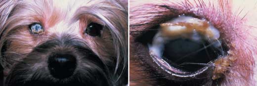
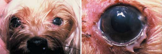
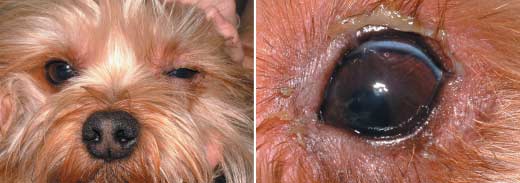
286 E T A L . Figure 1. Right eye of a 2-year-old, female Yorkshire Terrier with unilateral and severe keratoconjunctivitis sicca (STT 0 mm/min) and associated blepharospasm, blepharitis, copious mucoid discharge, and superficial corneal vascularization. Figure 2. Left eye of a 2-year-old, female Yorkshire Terrier with unilateral keratoconjunctivitis sicca (STT 4 mm/min) of 3 months duration. Blepharospasm, blepharitis, mucoid discharge, and superficial corneal vascularization can be observed. Figure 3. Left eye of an 11-month-old, male Yorkshire Terrier with unilateral and severe keratoconjunctivitis sicca (STT 0 mm/min) and associated blepharospasm and mild mucoid discharge.
affected than were male dogs (P = 0.05). No dogs were
the STT in the affected eye was 0 mm/min. Results of the
neutered and a familial relationship between the dogs was
STT in the unaffected eye ranged from 11 to 17 mm/min
known for only two dogs, which were mother and daughter.
(mean ± SD, 14 ± 2; median, 15). IOP was normal in all dogs.
The dogs ranged in age from 5 months to 4 years (mean ±
Affected eyes had blepharitis, mild conjunctival hyperemia,
standard deviation (SD): 2.1 ± 1.4 years; median: 2 years).
and variable degrees of superficial corneal vascularization
Duration of clinical signs before presentation ranged from 3
with a dry appearance to the ocular surface. Corneal melanosis
to 24 (mean ± SD: 9.1 ± 6.9; median: 6) months. All dogs
was not observed in any dog. The rest of the anterior segment
had been treated with various topical ophthalmic antibiotics
examination and the fundic examination revealed no
by the referring veterinarians, without resolution of clinical
abnormalities in any dog. Signs of cranial nerve dysfunction
signs. None of the dogs had been treated with sulfonamides
were not observed in any dog and no other relevant clinical
or had undergone surgery involving the third eyelid gland. A
signs were detected during the general physical examination.
history of trauma or infectious disease could not be obtained
Eleven dogs were initially treated with 0.2% cyclosporine
ophthalmic ointment (Optimmune, Schering Plough,
Thorough ophthalmic examination including the Schirmer
Buenos Aires, Argentina and Zaragoza, Spain) three times
tear test (STT) (Schering Plough Animal Heath Co., Kenil-
daily for 30 days. A mild reduction in the amount of mucoid
worth, NJ, USA), measurement of intraocular pressure
discharge and blepharospasm was observed in these eyes;
(IOP) using applanation tonometry (Tonopen, Mentor,
however, none of the treated eyes showed improvement
Norwell, MA, USA), application of fluorescein stain, slit-
in their STT values at revisit examinations during this time.
lamp biomicroscopy (Kowa SL-14, Kowa, Tokyo, Japan),
In fact, STT results declined by a further 3 mm/min and
and direct and indirect ophthalmoscopy was performed
4 mm/min, respectively, in the two dogs receiving this
in all dogs. Results of the STT in the affected eye ranged
treatment. The remaining five dogs that did not receive
from 0 to 7 mm/min (mean ± SD, 1 ± 2; median, 0). In 11 dogs,
cyclosporine, along with those three in which cyclosporine
2007 American College of Veterinary Ophthalmologists, Veterinary Ophthalmology, 10, 285–288
287
was unsuccessful, were treated twice daily with 20% chon-droitin sulfate ophthalmic solution (Tears, Labyes, BuenosAires, Argentina). All eyes receiving 20% chondroitin sulfateophthalmic solution showed subjective improvement inocular comfort as evidenced by reduced blepharospasm.
Open (lateral approach) parotid duct transposition (PDT)
was performed in three dogs, and a good result was obtainedin two. Excessive salivary secretion and facial dermatitisoccurred in the third. A nylon ligature was performedaround the duct to decrease salivary secretion in this dog.
History, signalment, clinical signs, age of presentation,
disease severity, and lack of response to medical treatmentsuggested that these dogs had breed-related unilateralaplasia or hypoplasia of the lacrimal gland. Based on thispresumptive diagnosis, biopsy of the lacrimal glands of theaffected eye was proposed in seven dogs but no owner con-sented to this. However, a 5-year-old, female dog, which hadundergone PDT, experienced acute spinal cord compressiondue to intervertebral disk protrusion and underwent reductionsurgery 2 years after the original ophthalmic diagnosis. Thisdog was subsequently euthanized owing to neurologic disease,and a tissue sample was collected from the site of the orbitallacrimal gland, fixed in paraformaldehyde and routinelyprocessed for histologic examination. Light microscopicexamination of a hematoxylin and eosin-stained section of
Figure 4. Photomicrograph of a tissue sample obtained from the
this tissue revealed absence of acini and ducts consistent with
dorsolateral orbit of a 5-year-old, female Yorkshire Terrier with
aplasia. A few lobules of sebaceous glands were observed
keratoconjunctivitis sicca and suspected congenital alacrima. Note the
(Fig. 4). Biopsy of the third eyelid lacrimal gland was not
absence of acini, ducts, inflammatory cells, or fibrosis consistent with
aplasia of the lacrimal gland. Vacuolated cells (arrows) represent
During the 4.5-year study period, only one other dog with
history, signalment, clinical signs, and response to therapysuggestive of congenital alacrima was presented to theauthors. This was a 1-year-old, female Chihuahua. This dog
well described.1,3,5 Aguirre et al. reported three cases of con-
was treated with 20% chondroitin sulfate ophthalmic solu-
genital KCS in a series of 71 dogs affected with dry eye.5
tion and showed a similar response to the Yorkshire Terriers
Although these authors described congenital KCS as more
common in miniature or small-breed dogs, there was no fulldescription of the clinical characteristics of these three dogs. In the current report, all but one of the affected dogs in a
DISCUSSION
4.5-year period were Yorkshire Terriers, suggesting that
Keratoconjunctivitis sicca is a common condition in dogs.
Yorkshire Terriers are predisposed to this syndrome. A sex
One of the most common causes of this syndrome is
predisposition for KCS has been suggested by some
immune-mediated destruction of the lacrimal glands, which
authors,1,3 and in other studies female were more commonly
tends to occur in adult dogs, is typically bilateral, and has an
affected than male dogs.10,11 In the study presented here,
insidious onset until aqueous deficiency is severe enough to
female (n = 12) were presented three times more commonly
produce signs of ocular surface dryness. The Yorkshire Terrier
than male dogs (n = 4) and this difference bordered on
is not a breed usually affected by the immune-mediated form
of KCS.1 The dogs in this study were all affected with severe
Congenital aplasia or hypoplasia of the lacrimal gland
clinical signs at an early age. Besides immune-mediated
should be considered when a young dog of a small breed is
dacryoadenitis, KCS may be caused by toxicity associated
presented with signs attributable to severe, unilateral reduc-
with sulfonamides,5,6 infectious disease, such as canine
tion in the aqueous layer of the precorneal tear film. This
distemper virus,4 facial and trigeminal nerve disorders,8 and
diagnosis is supported by a lack of history suggestive of
surgical excision of the third eyelid gland.7 Evidence of these
trauma, infectious disease, toxicity, or previous surgery of
causes was not found in any patient presented in the current
the third eyelid. All of the dogs presented here met these
criteria. Although confirmation of the diagnosis by biopsy of
Congenital alacrima, caused by aplasia or hypoplasia of
the lacrimal glands was not possible in most dogs in the pre-
the lacrimal gland, has been frequently mentioned but not
sent series because of lack of owner consent, histopathologic
2007 American College of Veterinary Ophthalmologists, Veterinary Ophthalmology, 10, 285–288 288 E T A L .
examination performed in one dog was consistent with
REFERENCES
lacrimal gland aplasia. Unfortunately, examination of the glandof the third eyelid was not performed in this dog. However,
1. Kaswan RL, Salisbury MA. A new perspective on canine kerato-
the extremely low STT values obtained in most affected eyes
conjunctivitis sicca. Treatment with ophthalmic cyclosporine. Veterinary
in the dogs presented here suggests that both lacrimal glands
Clinics of North America (Small Animal Practice) 1990; 20: 583 –613.
2. Kaswan RL, Bounous D, Hirsh SG. Diagnosis and management of
Treatment of these dogs was problematic, which is under-
keratoconjunctivitis sicca. Veterinary Medicine 1995; 90: 539–560.
standable if most had glandular aplasia or hypoplasia.
3. Moore CP. Diseases and surgery of the lacrimal secretory system.
In: Veterinary Ophthalmology, 3rd edn. (ed. Gelatt KN) Lippincott,
Cyclosporine is an immunosuppressive drug that produces
Williams & Wilkins, Philadelphia, 1999; 583–618.
immune modulation of the ocular surface and lacrimal
4. Martin CL, Kaswan RL. Distemper-associated keratoconjunctivitis
glands with associated increased tear production.1,3,11–13
sicca. Journal of the American Animal Hospital Association 1985; 21:
However, this therapeutic effect depends on the presence of
viable lacrimal gland tissue, and failure to effect an increase in
5. Aguirre GD, Rubin LD, Harvey CE. Keratoconjunctivitis sicca in
STT result in dogs with lacrimal gland aplasia or hypoplasia
dogs. Journal of the American Veterinary Medical Association 1971;
should be expected. Eleven of the dogs presented here were
158: 1566–1579.
6. Slatter DH, Blogg JR. Sulfonamide-related keratoconjunctivitis
initially treated using 0.2% cyclosporine ophthalmic
sicca in dogs. Australian Veterinary Journal 1978; 54: 444–446.
ointment but all failed to show improvement in STT values.
7. Morgan RV, Duddy JM, McClug K. Prolapse of the gland of the
Although some had mild reduction in mucoid discharge
third eyelid in dogs. A retrospective study of 89 cases (1980–1990).
and blepharospasm, this may have been due to the anti-
Journal of the American Animal Hospital Association 1993; 29: 56 –
inflammatory or lubricating properties of the drug. Cyclo-
sporine was not used at higher concentrations in these
8. Kern T, Erb HN. Facial neuropathy in dogs and cats: 95 cases
dogs because previous clinical trials performed by one of
(1973–1983). Journal of the American Veterinary Medical Association 1987; 191: 1604–1609.
the authors (HDH) showed no clinical differences in the
9. Sansom J, Barnett KC. Keratoconjunctivitis sicca in the dog: a
STT values of dogs with KCS treated with cyclosporine at
review of two hundred cases. Journal of Small Animal Practice 1985;
variable concentrations (data not published). 26: 121–131.
Chondroitin sulfate ophthalmic solution was used in three
10. Barnett KC. Keratoconjunctivitis sicca: sex incidence. Journal of
of the 11 dogs that failed to improve with cyclosporine, and
Small Animal Practice 1988; 29: 531–534.
as an initial treatment in five additional dogs. This drug
11. Lightowler CH, Herrera HD, Gómez NV. Lacrimomimetic effect
subjectively appeared to be more effective than cyclosporine
of topical cyclosporine A in canine KCS. Brazilian Journal of Veterinary Research and Animal Sciences 1993; 30: 233–241.
in reducing discomfort and mucoid discharge. This probably
12. Morgan RV, Abrams KL. Topical administration of cyclosporine
occurred as a result of its lacrimomimetic properties.3,14 The
for treatment of keratoconjunctivitis sicca in dogs. Journal of the
use of chondroitin sulfate as medical treatment of recurrent
American Veterinary Medical Association 1991; 199: 1043–1046.
corneal erosions has recently been described;15 however,
13. Olivero DK, Davidson MG, English RV et al. Clinical evaluation of
there is a paucity of references concerning the clinical use of
1% cyclosporine for topical treatment of keratoconjunctivitis sicca
tear substitutes like chondroitin sulfate and hyaluronic acid.
in dogs. Journal of the American Veterinary Medical Association 1991;
In addition, 20% chondroitin sulfate ophthalmic solution is
199: 1039–1042.
14. Herrera HD. Evaluación del uso tópico del ácido hialurónico como
highly viscous and would aid lubrication and protection of
humectante y regenerador del epitelio corneal en el canino. Revista
the ocular surface, reduce desiccation, and provide some
de Medicina Veterinaria 1997; 78: 46–50.
15. Ledbetter EC, Munger RJ, Ring RD et al. Efficacy of two chon-
Open PDT was performed in three dogs and was associated
droitin sulfate ophthalmic solutions in the therapy of spontaneous
with excessive salivary secretion and facial dermatitis in one
chronic corneal epithelial defects and ulcerative keratitis associated
of these. Other reported complications of this technique,
with bullous keratopathy in dogs. Veterinary Ophthalmology 2006; 9:
such as superficial band keratopathy caused by calcium
16. Barnett KC, Sansom J. Diagnosis and treatment of keratoconjunc-
precipitates or a failure to salivate,1,3,5,16,17 were not observed
tivitis sicca in the dog. Veterinary Record 1987; 120: 340–345.
in the dogs undergoing PDT in this study. The PDT
17. Gelatt KN. Treatment of canine keratoconjunctivitis sicca by
technique should be considered when no improvement in
parotid duct transposition. Journal of the American Animal Hospital
clinical signs is achieved with other therapies. Association 1970; 6: 1–12.
2007 American College of Veterinary Ophthalmologists, Veterinary Ophthalmology, 10, 285–288
Psychiatric and Behavioral Problems in Individuals with Intellectual Disability This checklist is based on Treatment of Psychiatric and Behavioral Problems in Individuals with Mental Retardation: An Update of the Expert Consensus Guidelines (2004) by M. C. Aman, M. L. Crismon, A. Frances, B. H. King, and J. Rojahn, which summarized the recommendations of a panel ofnational experts.
Performance Drug List For the most up-to-date Performance Drug List visit www.caremark.com The Caremark Performance Drug List is a guide within select therapeutic categories for clients, plan participants and health care providers. Generics should be considered the first line of prescribing. If there is no generic available, there may be more than one brand-name medicine to treat a condi
 286 E T A L .
 287
287